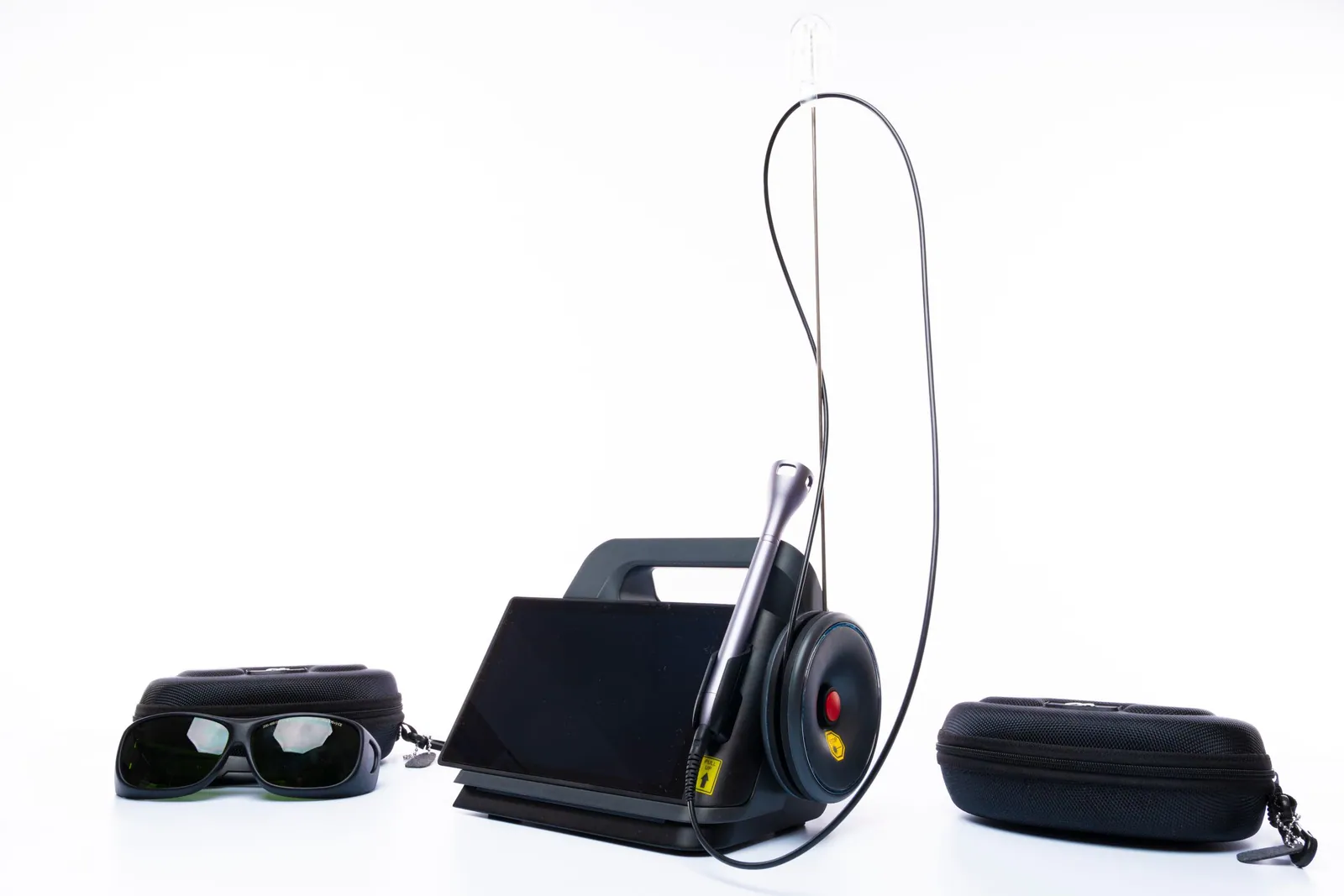
Dental lasers are fascinating, and the promise is appealing: treating the gums “without a scalpel,” disinfecting periodontal pockets with a beam of light, speeding up healing, reducing pain. Let us say it plainly from the outset: the laser is a real tool, sometimes useful in periodontics, but the scientific evidence remains nuanced. As things stand, professional societies regard it as a possible adjunct — an add-on — and not as a replacement for the standard of care, which remains the mechanical removal of tartar and biofilm beneath the gum (scaling and root planing).
As a dental surgeon in Kénitra, I offer here an educational, sourced overview of the laser in periodontics: how it works, what types of laser exist, what research actually shows about periodontitis, gum surgery and biostimulation, and above all what this technology does not deliver. This article explains the technology in general terms; it does not claim that any particular device is a miracle solution. Every clinical statement points to a verifiable source, and the benefits are presented as probabilities, never as a guarantee.
Laser for periodontal disease: the short answer
A laser for periodontal disease is used as an add-on to the deep cleaning of the roots, not instead of it. Added to root planing, the Er:YAG laser has shown a slight gain in the short term that then fades, and the evidence for the diode laser is thin. That is why the European Federation of Periodontology does not recommend the laser as an alternative to standard periodontal therapy.
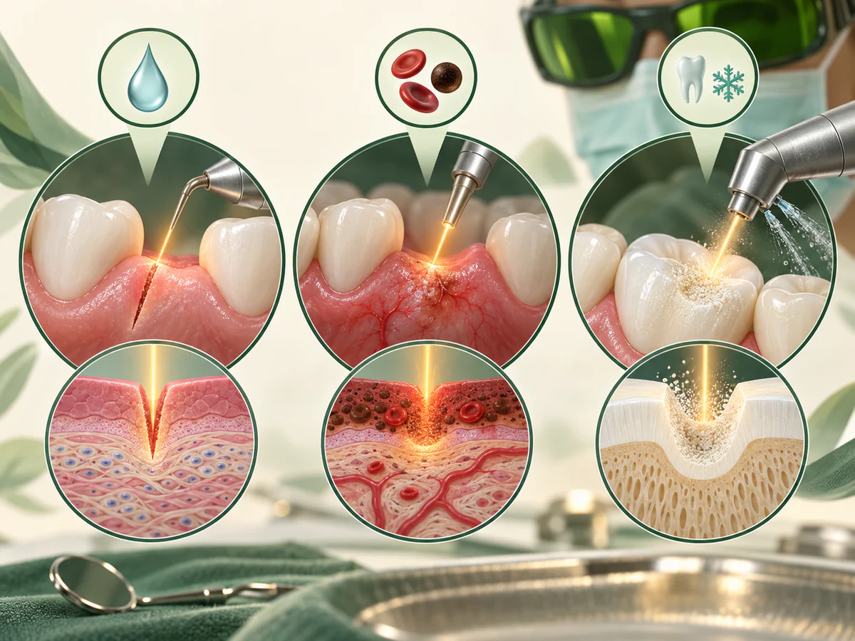
Lasers in periodontics have four main uses, each with its own level of evidence, detailed in the sections below:
- Inside the pockets, as an adjunct to root planing: a small, short-term benefit, if any.
- Gum surgery: cutting soft tissue with little bleeding, a more tangible advantage.
- Biostimulation: low-intensity light to support healing and ease pain, promising but not yet established.
- Around implants: inconsistent results, so an adjunct only.
How does a dental laser work on the gums?
The word “laser” refers to a concentrated beam of light at a precise wavelength. Depending on the target — the water in the tissues, bacterial pigments, melanin — this wavelength is absorbed to a greater or lesser degree, which produces different effects: cutting, vaporizing, disinfecting or stimulating healing. This is why there is no single “dental laser,” but several families, each with its own indications.
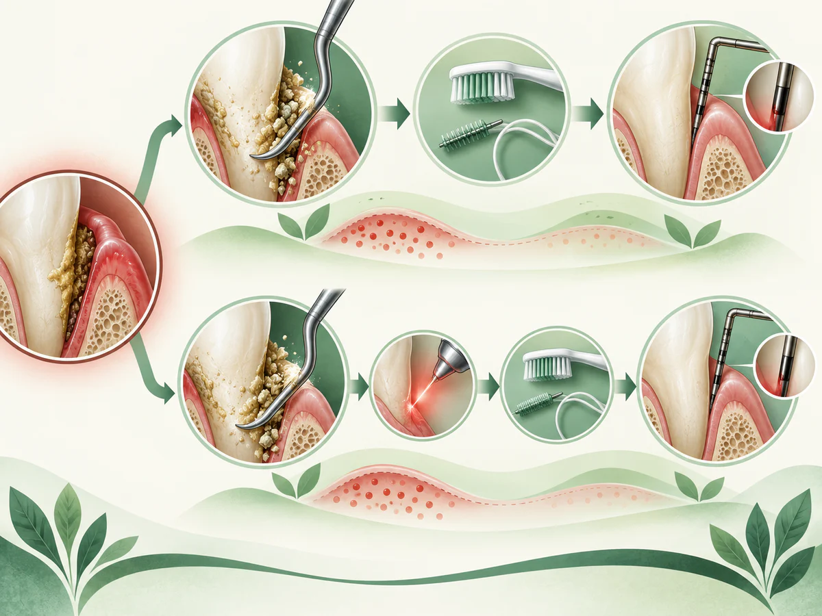
In periodontics, three main actions are expected of the laser. First, an antibacterial action: the heat of the beam can reduce the microbial load inside the periodontal pocket. Second, an action on the soft tissues: depending on the wavelength, the laser can cut or remove diseased gum tissue with little bleeding. Finally, an action known as biostimulation or photobiomodulation, at low intensity, intended to support healing and reduce pain. These three promises do not rest on the same level of evidence, and it is important to tell them apart.
The main types of laser in periodontics
Two large families come up repeatedly in the literature devoted to the gums.
The diode laser (wavelengths of roughly 810 to 980 nm) is the most widespread in practice, as it is compact and relatively affordable. It is absorbed mainly by pigments and acts on the soft tissues: it is used to decontaminate pockets, to remove granulation tissue, or for certain esthetic procedures on the gum. It does not act on tartar.
The Er:YAG laser (erbium, 2,940 nm) is strongly absorbed by water. It can act on both soft and hard tissues, and it is the only one that can, in theory, remove tartar from the root surface. That is why it has been the most studied as a possible alternative or complement to conventional instrumentation. The Nd:YAG laser is sometimes cited in one particular surgical protocol, but its evaluation remains limited.
The laser for treating periodontitis: what does the evidence say?
This is where caution is called for, because marketing often moves faster than science. The standard of care for periodontitis remains scaling and root planing — the subgingival instrumentation that mechanically disrupts and removes the bacterial biofilm. The real question is therefore not “does the laser work?” but “does the laser, added to this standard treatment, bring an additional benefit that is measurable and lasting?” And the answer, reading the data, is nuanced.
The Er:YAG laser: a short-term benefit that fades
The Er:YAG laser is the most promising on paper, since it can act on the root. The meta-analysis by Ma and colleagues (Medical Science Monitor, 2018), which pools ten randomized controlled trials, compared “Er:YAG laser + root planing” with root planing alone. At three months it observed a slight, statistically significant advantage for the laser: an additional pocket reduction of about 0.3 mm, a comparable attachment gain, and above all less pain reported. But — and this is the essential point — that advantage disappears at six and at twelve months: no significant difference remains between the two groups over the longer term.
An umbrella review published in BMC Oral Health (2024), which synthesizes several systematic reviews on the Er:YAG laser in non-surgical treatment, points in the same direction: the benefits, where they exist, are modest and heterogeneous, and the methodological quality of the studies remains limited. In other words, the Er:YAG laser may add a small something in the short term, particularly in terms of comfort, but it does not transform the outcome of treatment over time.
The diode laser: even thinner evidence
For the diode laser, the one most used in everyday practice, the data are less favorable. The review by Ren and colleagues, published in Evidence-Based Dentistry (2015), concludes that there is “little evidence” in favor of using the diode laser as an adjunct to non-surgical periodontal therapy: adding the laser did not convincingly improve clinical parameters compared with root planing alone. More recent individual studies sometimes suggest a short-term benefit with certain power settings, but the heterogeneity of the protocols makes any firm conclusion impossible. Biologically, the diode laser can reduce the bacterial load in the pocket, without this systematically translating into a better clinical result measured in millimeters.
Where the professional societies stand
The European Federation of Periodontology (EFP) has been clear on this point. In its S3-level clinical practice guideline on the treatment of stage I to III periodontitis (Sanz et al., Journal of Clinical Periodontology, 2020), it states that the laser is not recommended as an alternative to subgingival instrumentation, for lack of sufficient and consistent evidence. The available trials involved small numbers of participants and often carried an unclear risk of bias. The message is consistent with the one in our article on the difference between periodontitis and gingivitis: the foundation of treatment remains the mechanical removal of biofilm, whatever the favored tool of the moment.
| What the laser can bring | What the laser does not bring |
|---|---|
| A slight short-term gain with Er:YAG (≈ 0.3 mm) | A lasting superior result at 6–12 months |
| Less pain reported during and after the procedure | A replacement for scaling and root planing |
| A reduction in the bacterial load within the pocket | The rebuilding of bone already destroyed |
| Soft-tissue surgery with little bleeding | Any exemption from hygiene and maintenance |
| Possible help with healing (biostimulation) | A definitive cure for periodontitis |
The laser in gum surgery and for biostimulation
Beyond the periodontal pocket, the laser has other uses, where its value is sometimes more tangible — but always to be kept in perspective.
Soft-tissue surgery “without a scalpel”
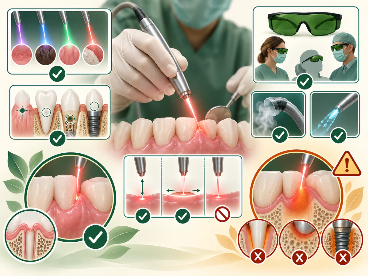
The laser, in particular the diode and the Er:YAG, makes it possible to carry out certain procedures on the gum while limiting bleeding, which improves visibility during surgery. Examples include removing granulation tissue, reshaping a gummy smile, treating certain overgrowths, or gingival depigmentation. The “without a scalpel” argument is partly real for the soft tissues: the beam coagulates as it cuts. That does not mean “without anesthesia” or “without after-effects”: local anesthesia is still needed in most cases, and healing takes time. For reconstructive periodontal procedures such as grafting in cases of gum recession, the laser does not replace the established surgical techniques.
Biostimulation and photobiomodulation
Photobiomodulation (low-intensity laser, sometimes called LLLT) neither cuts nor disinfects: it aims to stimulate the cells in order to reduce pain and support healing. The systematic review by Gopal and colleagues (Cureus, 2024) on the healing of oral wounds concludes that this approach has real potential to speed up healing and reduce postoperative pain, while stressing that larger studies, with genuine control groups, are needed to standardize protocols. A meta-analysis specific to gingival wound healing (Ebrahimi et al., BMC Oral Health, 2021) points the same way for post-surgical recovery: a plausible effect on pain and early healing, but a certainty that is still only moderate. So this is a promising field, not established proof.
And around implants?
The question comes up often: can the laser treat peri-implantitis, that infection around an implant? The available systematic reviews show inconsistent results: the laser can reduce inflammation and pocket depth around the implant, but without reliably outperforming mechanical debridement, and often with short follow-up. Here again, the laser is at best an adjunct. Prevention and rigorous follow-up remain decisive, as we explain in our article on periodontal maintenance and follow-up.
Laser or standard treatment: what changes in the session and on the quote
The laser does not replace standard treatment: when it is used, it is added to it.
Standard treatment of periodontitis follows a step-by-step path:
- An assessment. Probing measures the depth of the pockets tooth by tooth, and the X-ray shows the bone level.
- Control of plaque and risk factors. Learning to brush and clean between the teeth, professional scaling, and managing smoking or diabetes.
- Cleaning the roots beneath the gum. This is scaling and root planing, carried out under local anesthesia, often section by section.
- A reassessment, then maintenance. The pockets are checked again a few weeks later, and regular follow-up helps keep the result stable.
In the studies cited above, the laser is assessed as an addition to step 3: they compare root planing combined with a laser against root planing alone. It therefore removes no step from the path. In the session, it adds working time, with protective glasses for you and for the team, and the session does not become shorter.
The quote follows the same logic: a laser procedure is an additional item, not a treatment that replaces another. At the practice, gum treatment is quoted after the assessment, and the procedures involved and their fees are presented to you before anything begins. If a laser procedure is suggested to you, you can ask what it adds in your case and compare it with what the studies show: with the Er:YAG laser, a slight gain in comfort and pocket reduction in the short term, with no lasting difference at six or twelve months.
After laser periodontal therapy: caring for your gums at home
After laser periodontal therapy, the aftercare is the same as after root planing alone, because the laser is only an addition to it. Your gums may feel tender for a short while. Follow the instructions you are given, choose soft food if chewing is uncomfortable, keep brushing gently with a soft brush, treated areas included, and keep cleaning between your teeth as shown.
Contact the practice if swelling increases, if you develop a fever, or if bleeding does not stop. After that, the reassessment and maintenance visits matter more than the tool used: they show whether the pockets have stabilized.
What the laser does not bring: limits and precautions
Honesty requires naming the limits clearly, because they are often passed over in silence in commercial arguments.
- The laser does not replace mechanical removal of the biofilm. Root planing remains the foundation; the laser, at best, is added to it. A “laser-only” treatment is not supported by the guidelines.
- The laser does not rebuild lost bone. Like instrumentation, it can at best stabilize the disease; it does not spontaneously regenerate the support already destroyed by periodontitis.
- The demonstrated benefits are mainly short-term and small in magnitude. The difference of a few tenths of a millimeter observed with Er:YAG does not guarantee a better long-term outcome for the teeth.
- The settings matter enormously. Wavelength, power, application time: a poor setting can be ineffective, or even damage the tissues or the root surface. The operator’s skill matters more than the device.
- Safety is non-negotiable. The laser requires suitable protective eyewear for the patient and the team, and specific training.
- Neither the laser nor anything else removes the need for hygiene and maintenance. Without daily biofilm control and regular follow-up, no periodontal treatment holds up over time.
In short, the laser is one tool among others. In trained hands and for selected indications, it can bring comfort and a small benefit; it does not change the fundamentals of periodontal treatment, which rest on diagnosis, mechanical removal of the biofilm and serious follow-up.
In summary
The dental laser — above all the diode and the Er:YAG — is presented as a gentle way to treat the gums: antibacterial action, surgery with little bleeding, biostimulation, less pain. These strengths are partly real, but the evidence is nuanced. For periodontitis, the meta-analysis by Ma et al. (2018) shows a slight advantage for Er:YAG at three months that then fades, the evidence for the diode laser is thin (Ren et al., 2015), and the EFP (Sanz et al., 2020) does not recommend the laser as an alternative to subgingival instrumentation. Biostimulation is promising for healing and pain, without established certainty. The laser therefore remains a possible adjunct, never a replacement for scaling and root planing, and never removes the need for rigorous hygiene or regular maintenance.
Frequently asked questions
Can the laser replace scaling and root planing for treating periodontitis?
Is laser treatment on the gums really “painless” and “scalpel-free”?
Is the Er:YAG laser more effective than the diode laser for periodontitis?
Can the laser make lost bone or gum tissue grow back?
What is laser biostimulation or photobiomodulation?
Is the laser useful for treating an infection around an implant?
Sources
Medical references consulted for this article.
- 1Ma L, Zhang X, Ma Z, et al. Clinical Effectiveness of Er:YAG Lasers Adjunct to Scaling and Root Planing in Non-Surgical Treatment of Chronic Periodontitis: A Meta-Analysis of Randomized Controlled Trials. Med Sci Monit. 2018 (PubMed)
- 2Ren C, McGrath C, Jin L, Zhang C, Yang Y. Little evidence for the use of diode lasers as an adjunct to non-surgical periodontal therapy. Evid Based Dent. 2015 (PubMed)
- 3Sanz M, et al. Treatment of stage I–III periodontitis — The EFP S3 level clinical practice guideline. J Clin Periodontol. 2020 (Wiley Online Library)
- 4Effect of Er:YAG laser therapy in non-surgical periodontal treatment: an umbrella review. BMC Oral Health. 2024 (Springer Nature)
- 5Gopal L, Palwankar P, Dhalla N. The Efficacy of Low-Level Laser Therapy on the Healing of Oral Wounds: A Systematic Review. Cureus. 2024 (PMC)
- 6Effect of photobiomodulation in secondary intention gingival wound healing — a systematic review and meta-analysis. BMC Oral Health. 2021 (PMC)
- 7Clinical Evaluation of Adjunctive Nd:YAG or Diode Laser Application for Non-Surgical Periodontal Therapy: A Systematic Review with Meta-Analysis. Oral. 2026 (MDPI)
- 8Efficacy of non-surgical laser therapy for the management of peri-implantitis: A systematic review and meta-analysis. J Dent. 2025 (ScienceDirect)
Contents
Share