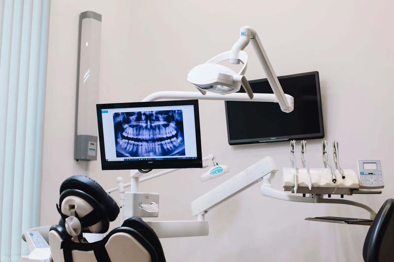
We hear more and more about “artificial intelligence” in the dental office, sometimes with overblown promises. The reality is simpler and more useful: there are now software programs that analyze your dental X-rays and flag suspicious areas for the clinician — an early cavity, a lesion at the tip of a root, bone loss around the teeth. This is often referred to as a “second look.”
That second look doesn’t decide anything for you, or for your dentist. It draws attention to details that are easy to miss on a small grayscale image, especially at the end of a long day. The diagnosis itself is always made by the clinician, who reads the X-ray alongside your clinical examination, your symptoms and your history. In this article, I’ll explain honestly what these tools can do, what they can’t, and why they remain an aid — never a replacement.
What is AI applied to dental X-rays?
Behind the term “artificial intelligence,” in this field, lies a specific technology: convolutional neural networks (CNNs). These are programs trained to recognize patterns in images. They have been shown tens of thousands of X-rays on which experienced dentists had marked, pixel by pixel, the exact location of cavities or lesions. Through repeated examples, the software learns to spot shapes and density variations associated with disease.
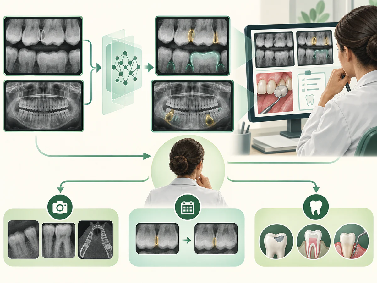
In practice, the clinician takes an X-ray as usual — a periapical film, a bitewing or a panoramic. The software analyzes it within seconds and overlays colored markers on the areas it considers suspicious, sometimes with a probability score. The dentist then reviews these suggestions and either confirms or dismisses them.
Two fundamental limits are worth stating right away. First, the software doesn’t “understand” anything: it recognizes statistical patterns, with no notion whatsoever of your pain or your history. Second, it depends entirely on the images it was trained on. If those images came from different equipment, a different population or a different quality level than your own film, its performance may drop. Researchers call this a generalization problem, and it is one of the reasons no tool should ever be used on autopilot.
It is important to understand what this is not. It is not a robot that “reads” your mouth, nor a system that makes a diagnosis on its own. It is an image-reading aid, in the same vein as those developed in general medical imaging. To better situate its role, it helps to understand the basics of 3D imaging and cone beam and of digital radiography, which this software relies on.
What AI helps to spot
Cavities, especially early ones
This is the most studied use. Cavities between the teeth (proximal cavities) are particularly hard to see with the naked eye and even on an X-ray, because they hide in the contact areas. Several systematic reviews have shown that algorithms achieve good detection performance. In the meta-analysis by Luke and Rezallah (2025, Head & Face Medicine), the weighted mean specificity approached 88%, while the authors also emphasized wide variability between studies.
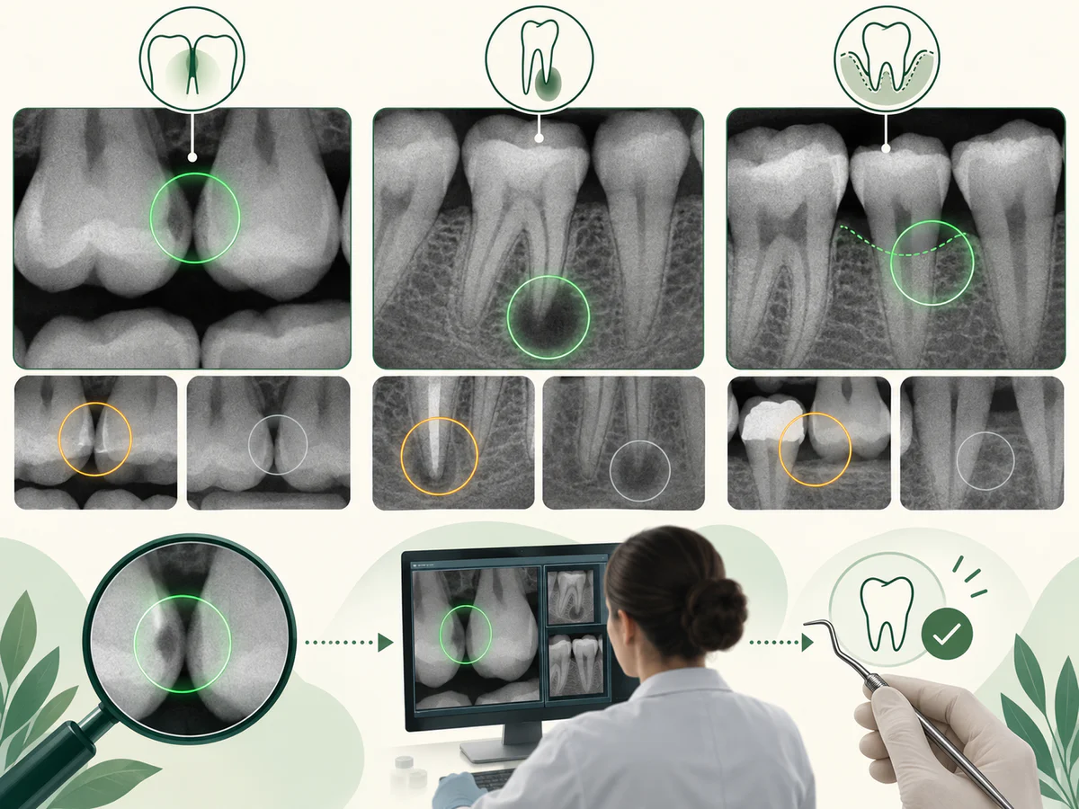
The main benefit is early detection. A cavity caught early can often be monitored or treated in a minimally invasive way, whereas a neglected cavity progresses into the dentin, and then toward the nerve. To understand that progression, you can read our article on treating cavities in adults.
A second benefit, less obvious to patients, is consistency. A dentist reads dozens of X-rays a day; fatigue, lighting or the pace of an appointment can cause a detail to be missed. Software, on the other hand, applies exactly the same level of attention to the first film of the day as to the last. It doesn’t replace the human eye, but it can act as a safety net against oversights caused by routine — provided the clinician remains the one who decides.
Lesions and bone loss
Beyond cavities, some software flags periapical lesions (at the tips of the roots) or measures the bone level around the teeth, a key indicator in periodontics. The systematic review by Khubrani and Thomas (2024, Dentomaxillofacial Radiology) reports, for the detection of periodontal bone loss on 2D X-rays, a pooled sensitivity of around 87% and a specificity of around 76%. Encouraging figures, but ones the authors qualify strongly, as we’ll see.
Monitoring bone level is central to managing periodontitis and gingivitis, where spotting early deterioration changes the prognosis.
Does AI really improve diagnosis?
That’s the right question. High performance “in the lab” does not guarantee a real benefit for the patient. Fortunately, clinical studies exist.
In a randomized trial published by Mertens et al. (2021, Journal of Dentistry), 22 dentists examined bitewing X-rays with and without software assistance. With AI support, their accuracy in detecting cavities was significantly better (area under the curve of 0.89 versus 0.85 without assistance). In other words, the tool helped them see more genuine lesions.
But — and this is essential — the same team showed, in a cost-effectiveness analysis (Schwendicke et al., 2022), that this increased accuracy could also lead to more decisions to treat invasively, with no demonstrated net gain in cost-effectiveness. Seeing more things does not mean everything should be treated. That is exactly where human judgment comes in.
| Aspect | What AI brings | What remains with the clinician |
|---|---|---|
| Reading the image | Flags suspicious areas within seconds | Confirming, dismissing, interpreting the context |
| Consistency | Doesn’t get tired, reads every film the same way | Cross-checking with the clinical exam and symptoms |
| Early detection | Helps spot subtle lesions | Deciding whether to monitor or treat |
| Treatment decision | None | Choosing the least invasive option suited to the patient |
What AI does not bring
This is the most important part of this article. A diagnostic aid is only useful if you know its limits.
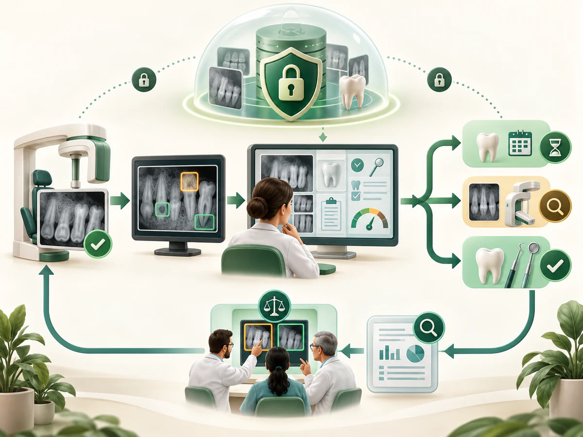
AI does not make a diagnosis. It flags probabilities on an image. A dental diagnosis takes in far more: your pain, your history, clinical tests, sometimes several films. A suspicious spot on an X-ray may be an artifact, an anatomical shadow or an old restoration. Only the clinician can tell the difference.
Performance varies a great deal from one software program and one study to another. Systematic reviews report substantial heterogeneity (I² values often above 86%, a sign that results diverge considerably). An impressive figure in one study is not a guarantee for every tool or for every patient.
False positives and false negatives do occur. No system is perfect. Software may flag a cavity that isn’t one (false positive), potentially prompting unnecessary treatment, or miss a genuine lesion (false negative), creating a false sense of security. Khubrani and Thomas (2024) in fact conclude that, in the absence of sufficient external clinical validation, these models “may not yet be good enough” as an automated screening tool.
The quality of the evidence still has room to improve. In that same review, fewer than a quarter of the studies reached a high level of quality, and more than half did not compare AI with clinicians in a blinded fashion. Research is advancing quickly, but rigorous multicenter trials are still needed.
The regulatory context matters. Several programs of this kind have obtained authorizations such as 510(k) clearance from the US FDA for detection assistance. This means they have been evaluated within a framework, but these authorizations cover decision support — explicitly not a replacement for the clinician.
And at the practice in Kénitra?
This article is deliberately educational: it explains a technology in general terms, so that you understand what the media and manufacturers are talking about. Whatever place AI holds in a given practice, the principle stays the same: the X-ray is a tool, and the decision belongs to the clinician who examines you.
At the practice, my priority is a considered, rigorous diagnosis that is as minimally invasive as possible. That means quality imaging, a careful clinical examination and a clear explanation of every option — whether it involves treating a cavity, managing periodontitis or planning an implant. If a lesion is spotted early, we can often favor monitoring or conservative treatment rather than a major procedure.
Honesty also requires saying this: no technology replaces good hygiene and regular follow-up. Careful daily dental hygiene and regular check-ups remain your best allies in avoiding, precisely, having to treat advanced lesions.
In summary
Artificial intelligence applied to dental X-rays is a real but modest aid: it offers a “second look” that helps the clinician spot cavities, lesions and bone loss earlier, with good performance in research settings. Clinical studies show it can improve detection — while reminding us that seeing more does not require treating more. Its limits are well documented: variability between software programs, false positives and false negatives, evidence quality that is still uneven, and a regulatory framework that defines it as an aid, never a substitute. The diagnosis, the clinical judgment and the treatment decision remain in the hands of your oral surgeon, who reads the image alongside your actual situation. It is in that partnership — a consistent tool and a cautious human judgment — that the real benefit for your oral health lies.
Frequently asked questions
Does artificial intelligence replace the dentist?
Is AI reliable for detecting cavities?
Can AI see bone loss or periodontitis?
Does AI mean more X-rays or more radiation?
Can an AI result be wrong?
Is seeing more lesions with AI always a benefit?
Sources
Medical references consulted for this article.
- 1Luke A.M., Rezallah N.N.F. (2025). Accuracy of artificial intelligence in caries detection: a systematic review and meta-analysis. Head & Face Medicine.
- 2Khubrani Y.H., Thomas D., et al. (2024). Detection of periodontal bone loss and periodontitis from 2D dental radiographs via machine learning and deep learning: systematic review employing APPRAISE-AI and meta-analysis. Dentomaxillofacial Radiology.
- 3Mertens S., Krois J., Cantu A.G., Arsiwala L.T., Schwendicke F. (2021). Artificial intelligence for caries detection: Randomized trial. Journal of Dentistry.
- 4Schwendicke F., et al. (2022). Cost-effectiveness of AI for caries detection: randomized trial. Journal of Dentistry.
- 5Cantu A.G., Gehrung S., Krois J., Chaurasia A., et al. (2020). Detecting caries lesions of different radiographic extension on bitewings using deep learning. Journal of Dentistry, 100:103425.
- 6Examining the diagnostic accuracy of artificial intelligence for detecting dental caries across a range of imaging modalities: an umbrella review with meta-analysis (2025). PLOS One.
- 7Artificial intelligence for radiographic imaging detection of caries lesions: a systematic review (2024). BMC Oral Health (PMC).
Contents
Share