In short: Dental microscopy is the use of an operating microscope that magnifies and lights the inside of the mouth, so the dentist can see details the naked eye cannot make out, such as a very fine canal or a crack. Its benefit is most clearly documented in endodontics, particularly in surgery at the tip of the root. It is a tool for precision, not a promise of results.
The dental operating microscope is an optical instrument that magnifies and brightly illuminates the working area, in order to make visible details that the naked eye cannot perceive: a very fine canal, a crack in the enamel, the exact border of a cavity or of a crown. Together with loupes, whose magnification is more modest, it makes up the family of vision aids in dentistry, grouped under the term “microscopic dentistry.” The central idea is simple: seeing better generally allows for more precise and more conservative work — that is, work that preserves more healthy tissue. It is in endodontics — the treatment of the inside of the root — that this contribution is best documented.
This article is a general explainer written for patients. It describes what magnification is, how it comes into play during treatment, and what the scientific literature says about it, without presenting the microscope as a guarantee of results or as equipment that is indispensable for every procedure. The aim is to give honest, nuanced information: magnification is a useful tool, sometimes a decisive one, but it replaces neither the diagnosis, nor the practitioner’s experience, nor the follow-up. The success of a treatment depends on many factors, and vision is only one of them.
What is magnification in dentistry?
The mouth is a cramped, dark and deep space, in which we work on structures only a few millimeters across. With the naked eye, and even with good lighting, certain details remain at the limit of what can be perceived. Optical aids address this problem by enlarging the image and by providing powerful light, most often along the line of sight (coaxial light), which removes the shadows at the bottom of the cavity. It is this combination of magnification and light that can enhance visual detail in such a small space.
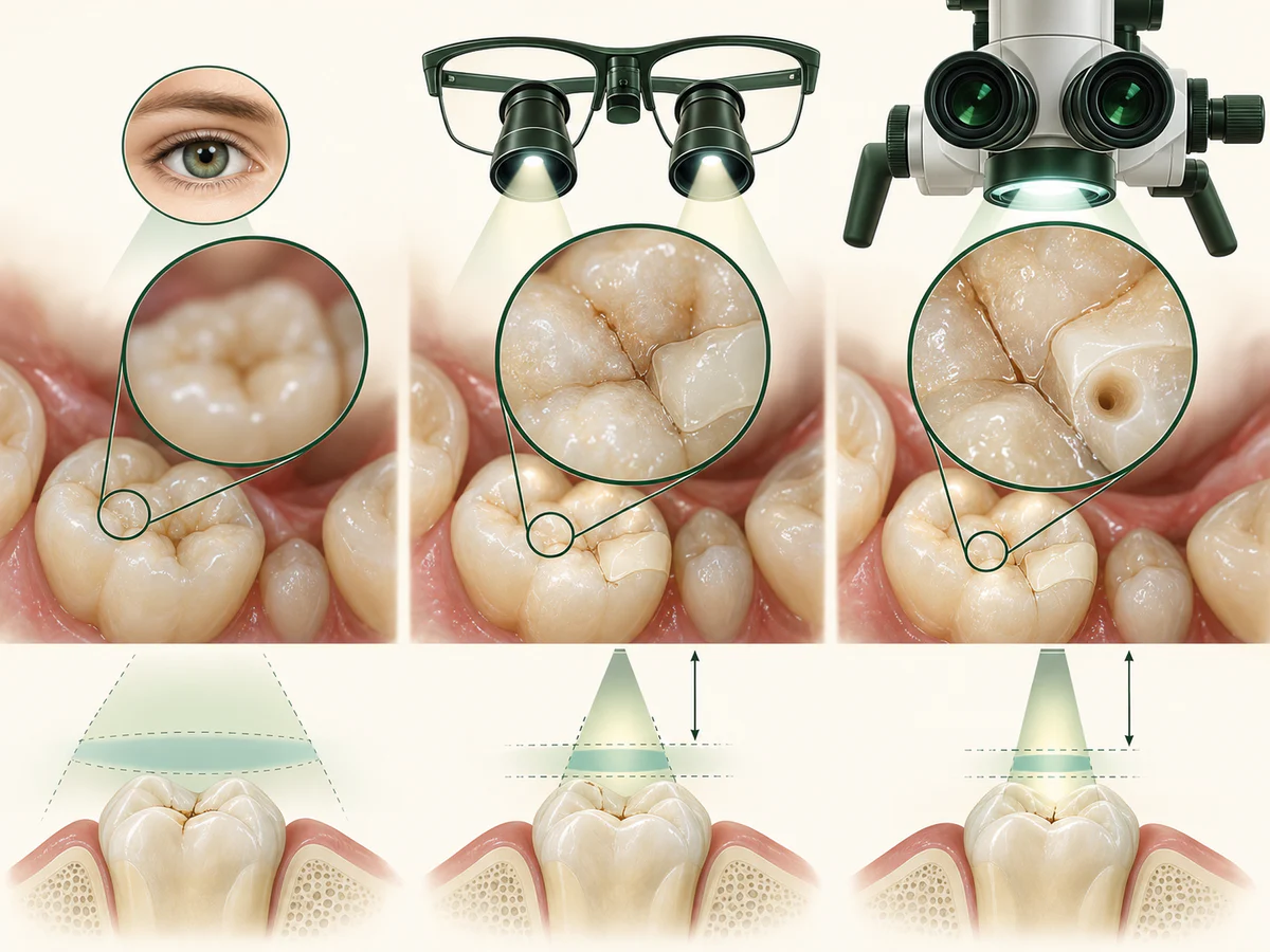
There are two main families of instruments, and they do not compete with each other — they complement one another.
Loupes
Loupes are magnifying systems mounted on eyeglasses. Their magnification generally ranges from 2.5 to about 6 times. They are light and mobile, and they are used for a large share of everyday procedures. They offer a first level of precision and, as we will see, an important benefit for the practitioner’s posture. For routine dental procedures such as fillings or scaling, this level of magnification is often sufficient.
The operating microscope
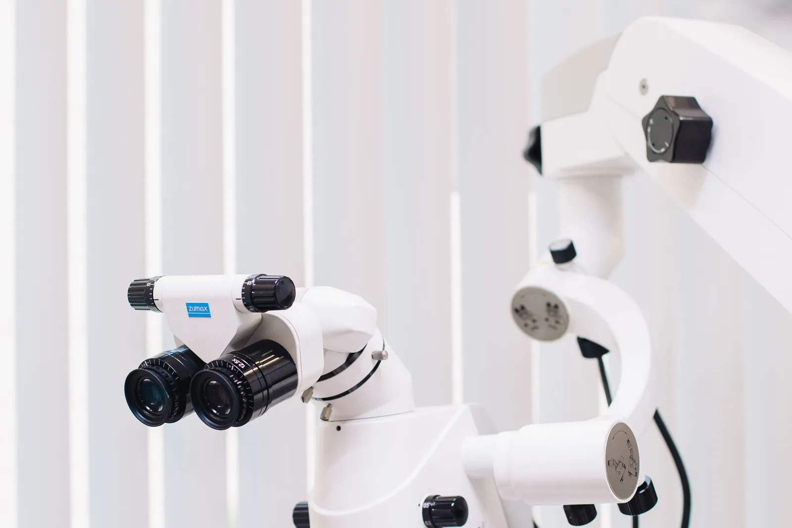
The dental operating microscope (or surgical operating microscope) is a more powerful device, mounted on an articulated arm, offering adjustable magnification that often ranges from 3 to more than 20 times, with intense coaxial lighting. It provides a stable and highly detailed image, at the cost of a more fixed working position and a longer learning period. According to the summary published by the American Association of Endodontists (Colleagues for Excellence, 2016), the microscope has transformed the practice of endodontics by making visible structures that were until then inaccessible to the naked eye.
How dental microscopy works
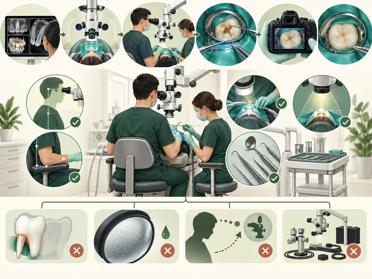
The dentist looks through two eyepieces, which give a three-dimensional view of the working area. A magnification changer lets the dentist move from a low setting, to see the whole tooth and its surroundings, to high magnification, to examine the entrance of a canal or a fine crack. The light is built into the head of the microscope and follows the line of sight, so the bottom of a cavity is lit without shadows.
The head is carried by an articulated arm, which keeps the image steady above the mouth. Many models can also hold a camera that records what the dentist sees. Working this way takes training: the dentist often views the teeth in a small mirror placed in the mouth and learns to coordinate precise hand movements with a magnified image.
Why does seeing better help in treating?
Seeing better is not an incidental comfort: it is a condition for precision. When a detail is visible, it can be treated; when it is not, it risks being missed. This principle plays out on several levels. It applies to many dental treatments, not only to root canals.
The first is conservative dentistry. Clearly seeing the boundary between healthy tissue and affected tissue makes it possible, in theory, to remove decay without sacrificing healthy enamel or dentin beyond what is necessary. This minimally invasive approach echoes the one described in Tooth decay in adults: from diagnosis to treatment, where preserving tissue is a central objective.
The second is the detection of details that are otherwise invisible: an additional root canal, a micro-crack, a root fracture, a deposit of tartar below the gum line, the precise margin of a crown preparation. The finer the resolution, the more such elements can be spotted and taken into account.
The third is the quality of checking: being able to verify during treatment how well a restoration fits or how clean a cavity is reduces the risk of imperfection. A clinical study by Wang et al. (2024), published in the American Journal of Translational Research, compared restorations carried out with and without a microscope and observed a better marginal fit — better-adapted edges — in the microscope-assisted group. This is a result on one specific technical criterion, measured in a single study, and not proof that all treatments performed under a microscope last longer.
Magnification in endodontics: where the evidence is strongest
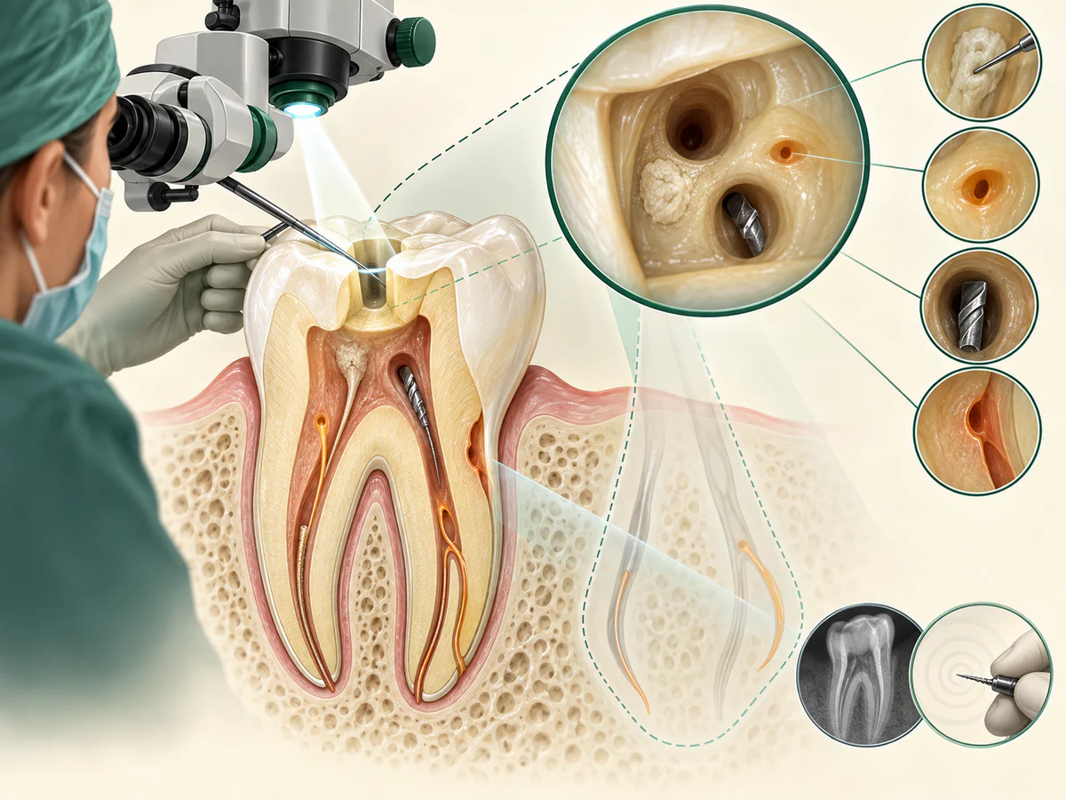
Endodontics consists of cleaning, disinfecting and filling the network of canals located inside the root. These canals are fine, sometimes curved, sometimes multiple and difficult to locate. It is one of the areas where vision makes a concrete difference, particularly in endodontic surgery (surgery on the tip of the root, or root-end resection).
The most frequently cited data comes from the meta-analysis by Setzer et al. (2012), published in the Journal of Endodontics. Comparing endodontic surgery performed with high magnification (microscope or endoscope) to a technique without high magnification, the authors found a significantly higher probability of success with high magnification. This analysis is often summarized as success rates that rise according to the vision aid used. The table below sets out these orders of magnitude drawn from the literature; they are averages from pooled studies, to be read as trends and not as an individual promise.
| Vision aid | Indicative magnification | Trend observed in endodontic surgery |
|---|---|---|
| Naked eye / no high magnification | 1x | Lowest success rate in pooled studies |
| Loupes | 2.5x to 6x | Intermediate level |
| Operating microscope | 3x to 20x and above | Highest reported success rates (Setzer et al., 2012) |
The reason for this benefit is straightforward: in endodontic microsurgery, seeing the tip of the root, spotting a missed canal or a crack, and checking the quality of the retrograde filling are all steps that magnification and light make feasible with precision.
A root canal treatment nevertheless remains a procedure in which how it unfolds and what follows matter as much as the technology used. The practical aspects of this treatment are covered in Root canal treatment: why, how, and the question of pain.
Root canals and diagnosis under the dental microscope
In root canal treatment, the challenge is often to find every canal. Root canals can be very narrow, partly blocked by calcification, or more numerous than expected, as with the extra canal often found in upper molars. High magnification and coaxial light make their entrances easier to locate. The microscope is also used when a treatment has to be redone, for example to remove old filling material or a fragment of a broken instrument from a canal.
Magnification also supports diagnosis and treatment planning. Under the microscope, the dentist can look for a fine crack in a tooth that hurts when biting, or check the edge of an old filling or crown for a gap. These findings are always combined with the clinical examination and the X-rays: the microscope shows more detail, but the diagnosis rests on the whole picture.
An often overlooked benefit: the practitioner’s ergonomics
Magnification does not only benefit the eyes; it also influences posture. Without an optical aid, the practitioner tends to lean forward and bend the neck to get closer to the mouth, a position that over time contributes to musculoskeletal disorders. And these disorders are very common in the profession: reviews on the subject report very high rates of neck, shoulder and back pain among dentists.
Loupes and the microscope, by optically bringing the image closer, make it possible to work with a straighter torso and a less tilted head. A review of postural ergonomics during periodontal treatment showed that wearing loupes improves several posture parameters, particularly at the head and neck. A 2024 study published in Scientific Reports also measured neck and shoulder muscle load during a crown preparation and observed a favorable effect of magnification aids on that load. So seeing better also helps the practitioner stay in the profession in better health — an indirect, but real, benefit for the quality of care.
What magnification does not provide
An honest presentation means stating the limits clearly. Magnification is a means, not an end, and on its own it does not transform an outcome.
It does not guarantee the success of a treatment. This is the most important point. The Cochrane review by Del Fabbro et al. (2015), devoted to magnification devices in endodontics, identified no randomized trial meeting its strict criteria that would allow the conclusion that magnification in itself improves the outcome of root canal treatment. Likewise, the systematic review by Del Fabbro and Taschieri (2010), in the Journal of Dentistry, found no significant difference in outcome between loupes, microscope and endoscope across the available studies. In other words: for endodontic surgery, high magnification is associated with better results (Setzer et al., 2012), but for conventional root canal treatment, the evidence of an advantage specific to the type of instrument remains limited. Success depends on a set of factors — diagnosis, disinfection, sealing, the condition of the tooth, follow-up — of which vision is only one component.
It does not replace skill or judgment. A microscope in insufficiently trained hands does not produce better care. The instrument reveals details; they still have to be interpreted and acted upon. The learning curve of the microscope, in particular, is real.
It is not indicated for everything, nor is it indispensable everywhere. Many high-quality treatments are carried out with the naked eye or with loupes. The presence of a microscope does not make a treatment better as a matter of principle, and its absence does not make it a poor one. The tool is justified when the fineness of the detail to be treated calls for it.
It is not a guarantee of durability or of freedom from complications. A better-fitting restoration has good reason to behave better, but no technology removes biological uncertainty, wear, or the need for rigorous hygiene and maintenance, as recalled in Everyday dental hygiene: the right habits.
What is a treatment under magnification like?
From the patient’s point of view, a treatment carried out with loupes or under a microscope looks a great deal like a conventional treatment. The main visible difference is the instrument: the practitioner wears loupe glasses, or positions above the working area a device on an articulated arm, topped with eyepieces and a light source.
The treatment is neither more painful nor systematically longer; in certain complex cases it may even gain in efficiency thanks to better visibility. The microscope also makes it possible to capture images or video of the treated area, useful for explaining to the patient what has been done and for documenting the file. This teaching dimension — showing rather than only describing — is one of the concrete contributions of microscopic dentistry.
In summary
Magnification, whether from loupes or from the operating microscope, helps in seeing what the naked eye cannot make out, and therefore in working with more precision and more respect for the tissues. Its benefit is best established in endodontic surgery, where the meta-analysis by Setzer et al. (2012) associates high magnification with better success rates, and it comes with a real ergonomic advantage for the practitioner.
But it is neither a guarantee of success nor a substitute for diagnosis, skill and follow-up. The reviews by Del Fabbro et al. (2010, 2015) remind us that the evidence of an advantage specific to the type of instrument remains limited for routine root canal treatment, and that the outcome of a treatment rests on multiple factors. Microscopic dentistry is a tool in the service of care: valuable where it counts, useful for patient education and for posture, but to be appreciated for what it is — an aid to vision, not a promise of results.
Frequently asked questions
What is the difference between loupes and a dental microscope?
Does the microscope make a treatment more painful or longer?
Does the microscope guarantee the success of a root canal treatment?
Is magnification useful outside of endodontics?
Should you choose a dentist equipped with a microscope?
Does magnification bring anything to the practitioner?
Can the dental microscope show me what the dentist sees?
Sources
Medical references consulted for this article.
- 1Setzer et coll. 2012, Outcome of Endodontic Surgery: A Meta-analysis of the Literature — Part 2: Comparison of Endodontic Microsurgical Techniques with and without the Use of Higher Magnification, Journal of Endodontics (ScienceDirect)
- 2Del Fabbro & Taschieri 2010, Endodontic therapy using magnification devices: a systematic review, Journal of Dentistry (PubMed)
- 3Del Fabbro et coll. 2015, Magnification devices for endodontic therapy, Cochrane Database of Systematic Reviews (PMC)
- 4Wang et coll. 2024, Impact of dental operative microscopes on precision in minimally invasive dental restoration procedures, American Journal of Translational Research (PMC)
- 5American Association of Endodontists, The Dental Operating Microscope in Endodontics, Colleagues for Excellence (Winter 2016)
- 6The effect of dental loupes on postural ergonomics during non-surgical periodontal therapy, 2025 (PMC)
- 7Effect of loupe and microscope on dentists’ neck and shoulder muscle workload during crown preparation, Scientific Reports 2024
Contents
Share