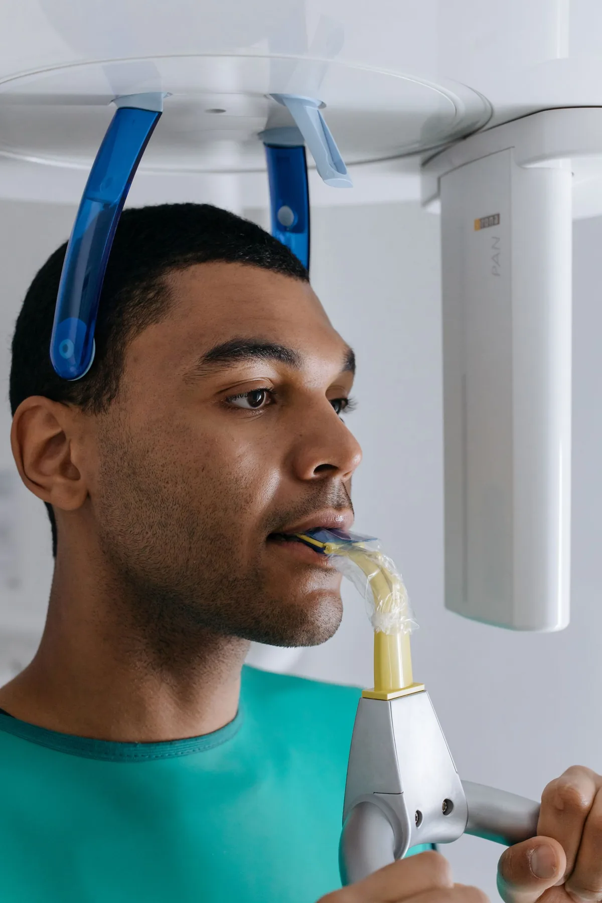
In short: A pano X-ray, short for dental panoramic X-ray, is a single image of the upper and lower jaws, taken by a machine whose rotating arm turns around your head for a few seconds. It gives an overview of the teeth, the jaw bone and nearby structures, with low radiation exposure. For fine detail of one tooth, a small intraoral film remains more precise, and a cone beam scan is used when a 3D measurement is needed.
The dental panoramic X-ray, also called an orthopantomogram, is a radiograph that shows the whole mouth on a single image: both arches, every tooth from right to left, the jaw bone, the joints and part of the sinuses. It is an overview examination, simple and quick, often prescribed to take stock of the mouth as a whole, to locate wisdom teeth or to prepare a treatment. But like any radiograph, it involves exposure to X-rays, it has specific indications, and it does not answer every question. This article explains what a panoramic X-ray really is, how it differs from a small “periapical” film and from a 3D Cone Beam, how the question of dose is approached, in which situations it is useful, and above all what it does not provide.
The aim is not to present the panoramic X-ray as essential at every appointment, nor as an image that “sees everything”. It is one diagnostic tool among others: it informs a decision, it does not treat anything and it guarantees no result. Honest information means saying both what it allows us to see and what it leaves in shadow, and recalling that ordering one follows rules of justification grounded in international recommendations.
What is a dental panoramic X-ray?
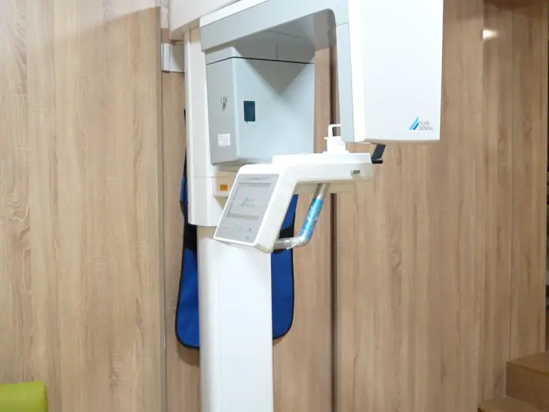
The panoramic X-ray is what is known as an “extraoral” radiograph: the machine stays outside the mouth. An X-ray source and a sensor rotate together around the head for a few seconds, and the image is built up as they move. The result is a single long, horizontal image that “unfolds” both jaws in two dimensions. The technique itself is called panoramic radiography, or orthopantomography.
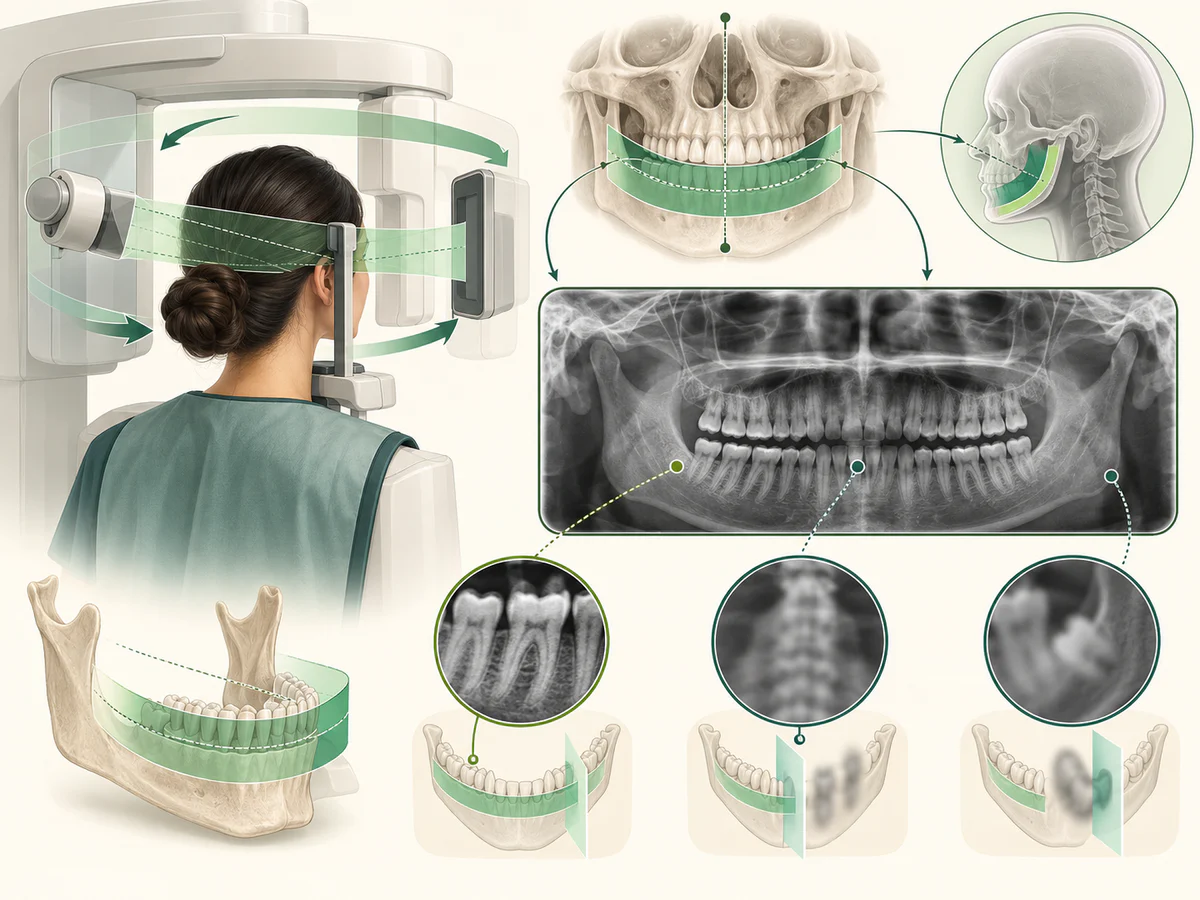
In practice, the examination is brief. You remove jewelry, glasses and removable appliances, stand or sit in the machine, bite gently on a small support to steady the head, and stay still for five to ten seconds while it rotates. Nothing to swallow, nothing to hold in the mouth for long: it is often more comfortable than small intraoral films for people with a strong gag reflex. The digital image is available immediately.
One inherent limitation should be understood, however: the panoramic X-ray is a two-dimensional image. It superimposes on a single plane structures that lie at different depths, and it slightly distorts volumes. It gives an excellent overview, but it does not precisely measure the thickness of the bone or the exact position of a nerve in space. For that, a 3-D image is needed, which is what a Cone Beam provides.
What a standard panoramic X-ray of the teeth shows
A standard panoramic X-ray shows all your teeth on one image, upper and lower, including teeth still hidden in the bone, such as wisdom teeth or, in children, adult teeth that have not yet come through. Around them, it shows the roots, the jaw bone, both jaw joints and the lower part of the sinuses.
On the image, dense materials look white: enamel, fillings, crowns and implants. Less dense areas look darker, which is why a large cavity or an area of bone loss can stand out. In the lower jaw, the canal that carries the nerve is often visible, but a flat image cannot show its exact depth. The word “standard” simply means the usual full-mouth view: some machines also offer shorter programs that cover only one area, such as the jaw joints, when only that area needs checking.
How the pano X-ray machine works
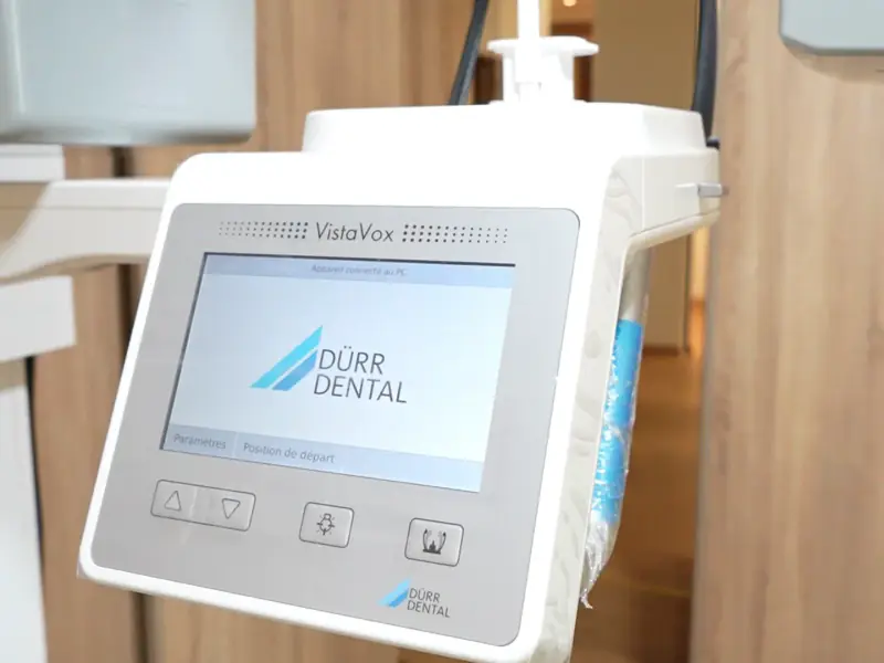
A panoramic X-ray machine consists of an X-ray source and a sensor fixed at opposite ends of a rotating arm. During the exposure, the source passes behind your head while the sensor moves in front of your face. Only a curved layer that follows the shape of the dental arches comes out sharp; structures in front of or behind it are blurred. This is how a single image can capture the entire mouth, from one jaw joint to the other. Some machines also carry an extra arm for cephalometric (“ceph”) X-rays, a side view of the skull used in orthodontic planning.
Positioning affects the quality of the image. Light guides help align your head, and you are asked to press your tongue against the roof of your mouth, because the air left there can show up as a dark band over the upper teeth. With digital panoramic X-rays, the image can then be enlarged and its contrast adjusted on screen, and it is stored in your file for comparison with later images. Because these are digitized images, they can also be shared with another practitioner if a referral is needed.
Digital panoramic X-ray or panoramic film?
Older machines recorded the image on dental panoramic film: a large sheet held in a cassette, then developed with chemicals before it could be read. Most panoramic X-ray machines now use a digital sensor instead, and the image appears on the screen moments after the exposure. For you, the examination is the same: same position, same rotating arm, same few seconds of keeping still.
Many people still say “panoramic film” for the image itself, even when it is digital. What a digital panoramic X-ray mainly changes is what happens after the exposure: no chemical development, and an image that can be adjusted on screen and compared with earlier ones. The dose depends above all on the machine and its settings, so the same rules of justification, described below, apply in both cases.
Panoramic, periapical and Cone Beam: what are the differences?
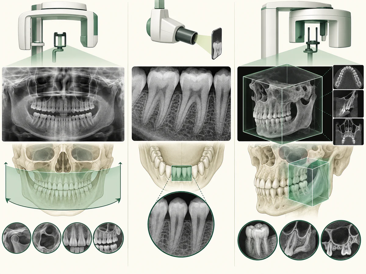
These three examinations are often confused. They do not show the same thing and they do not have the same indications. The periapical radiograph (an intraoral film) is a small, highly detailed image centered on one or a few teeth: it is the reference examination for closely analyzing decay between two teeth, the tip of a root or a site of infection. The panoramic X-ray, for its part, offers a wide but less detailed view of both arches. The Cone Beam (CBCT), finally, is three-dimensional imaging: it reconstructs a volume that can be explored in every direction, something no 2D radiograph allows.
These examinations are not in competition but complementary. The recommendations of the American Dental Association and the American Academy of Oral and Maxillofacial Radiology place the clinical assessment and 2D radiography as the first step, with 3D imaging coming in only when a specific question remains unanswered. The table below summarizes these differences, without any absolute hierarchy: each examination has its own indication.
| Examination | Dimension | What it shows well | Radiation dose |
|---|---|---|---|
| Periapical | 2D | One or a few teeth in detail: decay, root, apical infection | Very low |
| Panoramic | 2D | Overview of both arches, wisdom teeth, general assessment | Low |
| Cone Beam (CBCT) | 3D | Bone volume, position of nerves and sinuses | Higher, varies with the machine |
Each of these examinations is covered in detail elsewhere: how sensors and filmless radiography work in Digital radiography in the dental office, and 3D imaging in The Cone Beam (CBCT): 3D imaging at the practice.
The PA X-ray in dentistry: how it differs from a pano
In dentistry, “PA” stands for periapical. A PA X-ray is a traditional intraoral X-ray: a small sensor or film is placed inside the mouth, behind the teeth being examined. The image shows one to a few teeth in full, from the crown to the tip of the root, with the surrounding bone, in much finer detail than a panoramic image. It is used, for example, to examine an infection at the root tip or to check a root canal treatment.
A pano trades that detail for coverage: it shows the whole mouth at once, with less sharpness and some distortion. The two are complementary in treatment planning: the pano gives the overview, and a PA image answers a precise question about one area. Bitewings, another kind of small intraoral film, show the crowns of the upper and lower back teeth together and are mainly used to look for decay between teeth.
The question of dose: justification and optimization
The panoramic X-ray delivers X-rays, but at a dose that remains low. The review of doses in dental imaging published by Lee and Badal (2010-2020), available open access, reports an average effective dose of about 18 µSv for a panoramic X-ray (in the region of 3 to 75 µSv depending on the machine), compared with about 1 µSv for a periapical film and around 120 µSv for a CBCT. In other words, a panoramic X-ray represents roughly the exposure of a few days of natural background radiation: we are in fact permanently receiving “background radioactivity” of natural origin, which the World Health Organization (WHO) fact sheet notes makes up the bulk of our annual exposure.
That the dose is low does not mean it is negligible, nor that a panoramic X-ray is justified in every circumstance. Prescribing one follows two principles inherited from radiation protection.
The principle of justification holds that an examination should only be carried out if the expected diagnostic benefit outweighs the risk associated with the radiation. The WHO sums this principle up as follows: any decision that alters a person’s exposure to radiation should do more good than harm. A “routine” panoramic X-ray, with no warning sign and no clinical question, therefore does not meet this rule.
The principle of optimization consists of delivering the lowest dose compatible with an image that is useful for diagnosis: this is the ALARA principle (“as low as reasonably achievable”). This requirement is particularly important in children, who are more sensitive to radiation, and for whom the FDA and the American Dental Association recommend imaging only when necessary and adjusting the settings. Justification is always assessed case by case, after a clinical examination. The same reasoning guides the type of X-ray: the dental radiographic examination chosen is the one that answers the clinical question with the lowest dose compatible with a useful image.
In which situations is a panoramic X-ray useful?
The panoramic X-ray becomes valuable whenever an overview provides information that small films cannot give. Several indications are well established, always within the framework of a justified prescription.
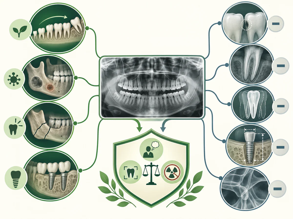
- Wisdom teeth. The panoramic X-ray is the first-line examination for viewing the position of the wisdom teeth, spotting an impacted tooth and assessing its relationship with neighboring structures before deciding on an extraction.
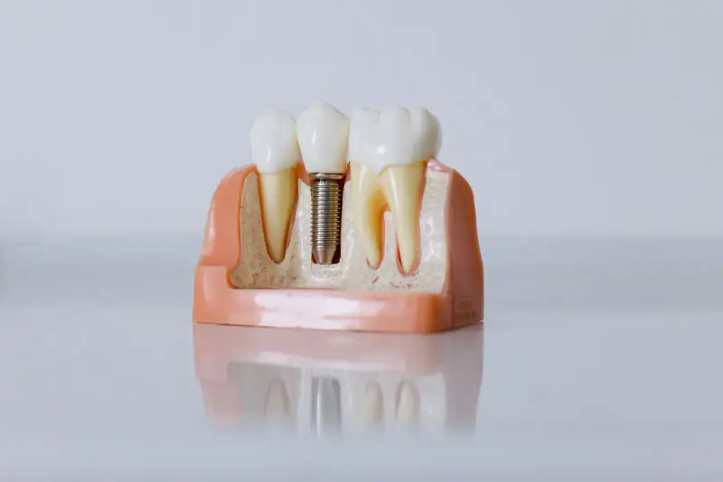
- Assessment before an implant. Before an implant is placed, the panoramic X-ray serves as the initial examination for a first evaluation of the bone and the overall context; when a precise measurement of bone volume is needed, it is supplemented by a CBCT. The full sequence is set out in our step-by-step guide to dental implant placement.
- Overall assessment. Cysts, impacted teeth, the condition of the bone around the teeth, developmental anomalies: the panoramic X-ray helps to map the situation as a whole, for example during an initial orthodontic assessment or a general review. It also shows the lower part of the maxillary sinuses, above the upper back teeth, and can reveal bone abnormalities that call for a closer look.
- Follow-up in children. As the permanent teeth come through, it can help check the presence and position of the tooth germs when the clinical examination justifies it.
What the panoramic X-ray does not provide
An honest presentation of this examination also means stating its limits. The panoramic X-ray is an overview tool, not an image that “sees everything”.
It detects small cavities poorly, particularly between the teeth. A comparative study by Kamburoğlu et al. (2012) showed that intraoral bitewing radiography is superior to the panoramic X-ray for diagnosing decay in the back teeth. For early decay, the panoramic X-ray therefore does not replace small targeted films; the diagnosis and treatment of decay are detailed in Tooth decay in adults.
It does not give the detail of a single tooth. To closely analyze the tip of a root, follow up a root treatment (root canal) or investigate localised pain, the periapical film remains more precise.
It does not measure in three dimensions. Because it flattens the relief, the panoramic X-ray does not reliably measure the thickness of the bone or the exact position of a nerve. When that information determines a surgical procedure, the CBCT takes over. This is typically the case when dental implants are planned close to a nerve or a sinus.
It is not a screening test for oral cancer. Changes in the lining of the mouth, the tongue or the gums are looked for during the clinical examination, by sight and touch.
Finally, it guarantees no result. An image, even a good-quality one, informs a decision: it removes neither the uncertainties of a treatment nor the need for a clinical examination and follow-up. The panoramic X-ray helps to understand a situation; it does not resolve it on its own. Your oral health is assessed on the whole picture: what the dentist sees in the mouth, your symptoms and, when they are justified, the images.
In summary
The dental panoramic X-ray is an overview radiograph — simple, quick and well tolerated — that shows both arches on a single two-dimensional image. It is useful for wisdom teeth, the initial assessment before an implant, general assessment and follow-up in children, at a low radiation dose, in the region of a few days of natural background radiation.
But it is neither a routine examination nor an image that sees everything. It detects small cavities poorly, does not give the detail of an individual tooth and does not measure in three dimensions: depending on the question being asked, the periapical film or the Cone Beam remain more suitable. Carrying one out follows two principles — justification, which reserves it for cases where the benefit outweighs the risk linked to the radiation, and optimization, which aims for the lowest dose compatible with a reliable diagnosis. At Dr Fatima Azelmat’s practice in Kénitra, imaging is used within this framework: a tool prescribed when it is useful, in the service of a considered decision.
Frequently asked questions
What is the difference between a dental panoramic X-ray and a small radiograph?
Is a dental panoramic X-ray dangerous? What radiation dose does it involve?
Does a panoramic X-ray show cavities?
Do you need a panoramic X-ray or a Cone Beam before an implant?
Is the dental panoramic X-ray suitable for pregnant women and children?
How often should you have a pano X-ray?
Is a “dental panorama” the same as a dental panoramic radiograph?
Sources
Medical references consulted for this article.
- 1FDA & American Dental Association, ADA/FDA Guide to Patient Selection for Dental Radiographs (justification, dose minimale, sensibilité des enfants)
- 2Lee et Badal 2021, A Review of Doses for Dental Imaging in 2010–2020 and Development of a Web Dose Calculator, Radiology Research and Practice (PMC, libre accès)
- 3Kamburoğlu et coll. 2012, Proximal caries detection accuracy using intraoral bitewing, extraoral bitewing and panoramic radiography, Dentomaxillofac Radiol (PubMed)
- 4OMS, Ionizing radiation and health effects (aide-mémoire, 27 juillet 2023)
- 5OMS, Justification and optimization in medical imaging (principes de radioprotection)
- 6American Dental Association & AAOMR 2025, Patient selection for dental radiography and cone-beam computed tomography, JADA
Contents
Share