In short: A cone beam CT (CBCT) is a dental X-ray examination that produces a three-dimensional image of the teeth, jaws, and surrounding structures, which can be explored in sections and measured. It plays an important role in implant planning and is also used for some impacted teeth and complex root canal cases; the scan itself usually takes just a few seconds. Because its dose is generally higher than that of conventional dental X-rays, it is performed only when the expected diagnostic benefit outweighs the radiation risk.
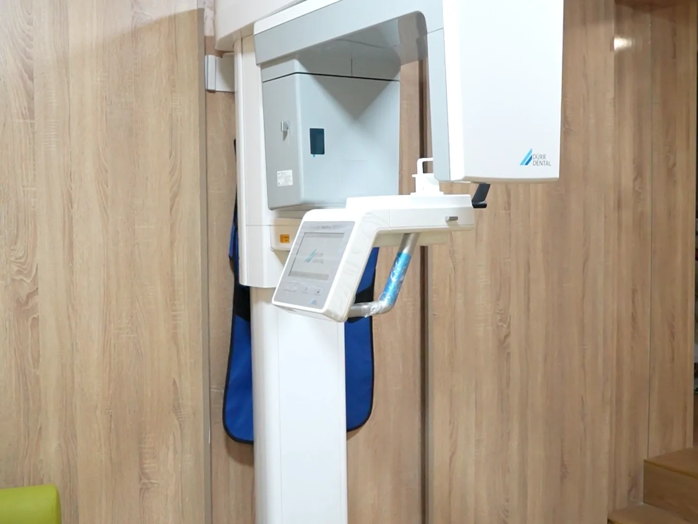
Cone beam imaging, or CBCT (cone beam computed tomography), is a radiological examination that produces a three-dimensional image of the teeth, jaws, and surrounding structures. Unlike a conventional X-ray, which projects depth onto a flat plane, CBCT reconstructs a volume that can be explored in every direction: height, width, depth. At our Kenitra practice, this examination is performed with a Dürr Dental Cone Beam unit. It is a diagnostic and planning tool, not a treatment in itself: it helps guide decisions, it does not guarantee any outcome. This article explains what CBCT is, how it differs from a 2D X-ray, what role it plays in the implant work-up, how the question of radiation dose is handled, and above all what it does not provide.
The goal here is not to present 3D imaging as essential to every consultation, nor as a guarantee of absolute precision. CBCT is a useful examination — sometimes a decisive one — but it involves exposure to X-rays and is not indicated in every situation. Honest information means explaining both what it can and cannot do, and remembering that its use is governed by strict justification rules based on international recommendations.
What Is a Cone Beam CT (CBCT)?
CBCT is a cone beam volumetric tomography device. During the examination, an X-ray source and a sensor rotate around the patient’s head, acquiring a series of images. A computer then reconstructs a three-dimensional volume that the practitioner can slice through and examine plane by plane. The examination is quick; the patient remains seated or standing, holding still, usually for just a few seconds.
CBCT should not be confused with a conventional medical CT scan (computed tomography). Both produce 3D images, but the technology differs: CBCT is designed for the dental and maxillofacial region, with fine resolution of bone and teeth, and it generally delivers a lower radiation dose than a medical CT scan. In the United States, the Food and Drug Administration (FDA) notes in its information sheet on Dental Cone-beam Computed Tomography that the dose from a dental CBCT is generally lower than that of other CT examinations, but higher than that of conventional dental X-rays.
How Does CBCT Differ from a 2D X-ray or a Panoramic?
The essential difference lies in the move from two dimensions to three. A periapical X-ray or a panoramic (orthopantomogram) are two-dimensional images: they superimpose structures located at different depths onto a single plane. They are informative about the presence of decay, an infection site, or the overall condition of the teeth, but they cannot measure bone thickness or the exact position of a nerve relative to a future implant site.
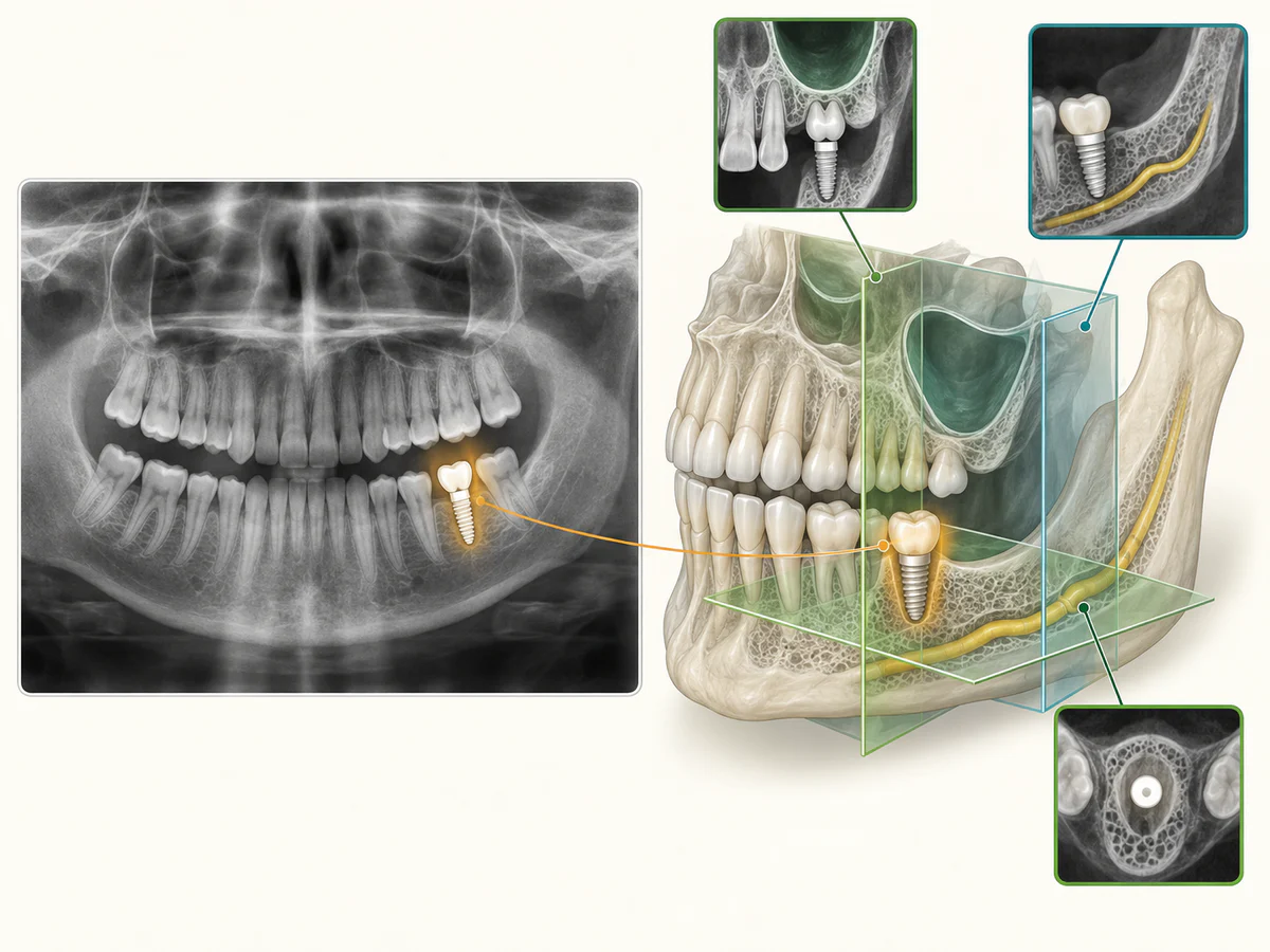
CBCT, by contrast, captures the full volume. It makes it possible to measure the height and width of bone available at a specific spot, and to locate in space the structures that must be protected. It is this three-dimensional information that gives it its value in certain surgical situations.
Imaging selection depends on the indication: routine consultation, implant planning and complex endodontics have different needs. The clinician reviews the case and previous images. The table compares uses without imposing a universal 2D-then-3D sequence.
| Examination | Dimension | What it shows well | Radiation dose |
|---|---|---|---|
| Periapical X-ray | 2D | One or a few teeth, decay, apical infection | Very low |
| Panoramic | 2D | Overview of both arches, global picture | Low |
| CBCT (Cone Beam) | 3D | Bone volume, position of nerves and sinuses | Higher than 2D X-rays, varies by device |
Understanding the images: volume, sections and measurements
Reconstructed volumes, radiographic sections and surface impressions answer different questions. The screen photograph below illustrates displayed views, not a complete case or a diagnostic demonstration.
01. Reconstructed volume: orientation
A 3D reconstruction can provide an overview. A visually clear rendering does not prove that every clinical question has been answered.
02. Cross-sections: explore an area
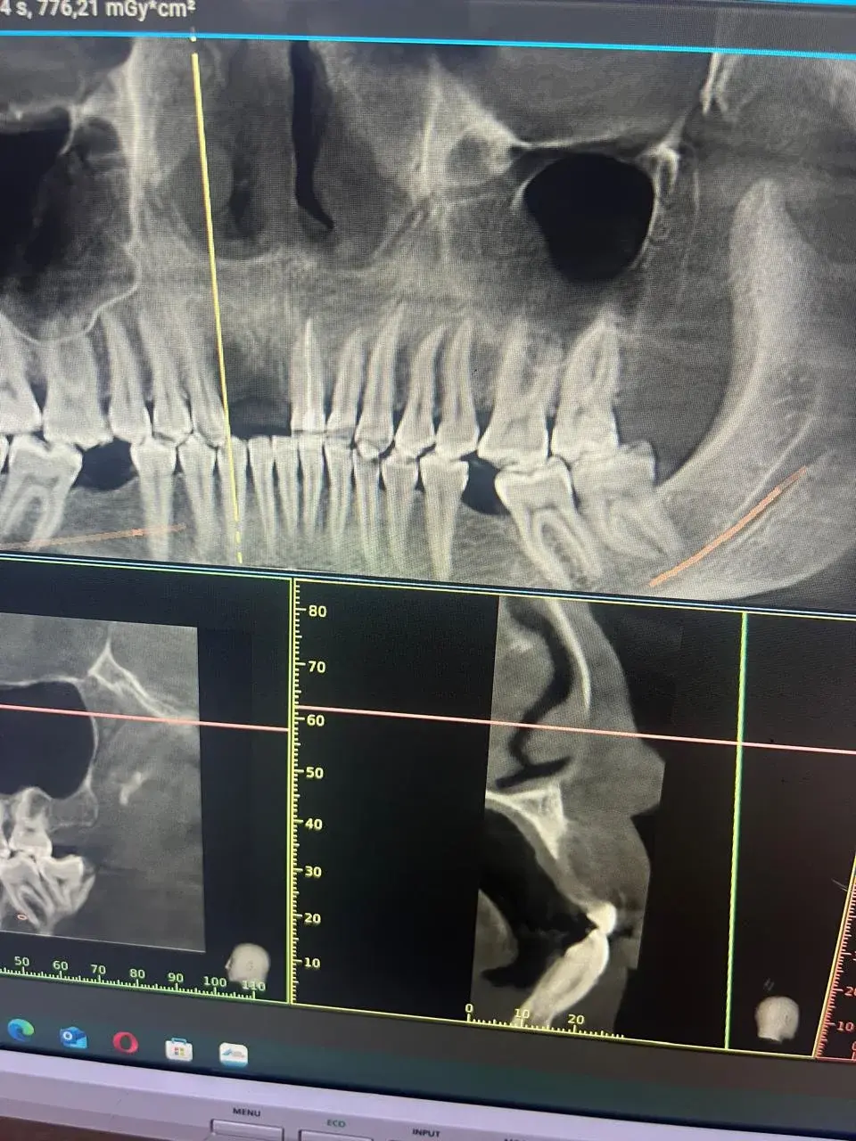
The clinician examines the planes needed for the clinical question. A panoramic image may appear alongside sections; this photograph alone does not identify its acquisition method.
03. Measurements: interpret them in the case context
Interpretation considers the complete examination and image quality. An isolated measurement is not a diagnosis, prescription or safety guarantee.
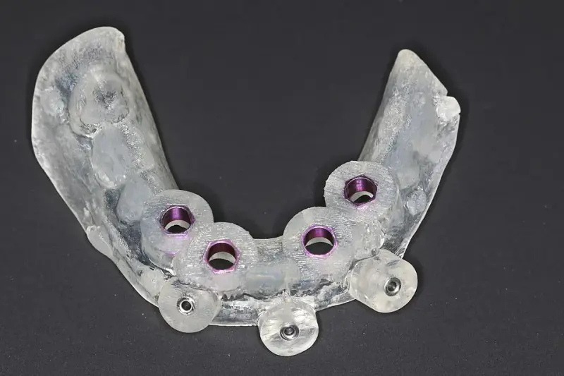
What about digital impressions? They record oral surfaces for prosthetic work, not the inside of bone as radiological imaging does. See the dental implant placement steps.
Before the scan: three useful questions
- Which clinical question should 3D imaging answer in my case?
- Can previous examinations or another imaging method provide the information?
- Which field of view and settings suit the area being examined?
The FDA explains that CBCT uses X-rays and should be justified and optimised. These general principles are not an individual prescription or Moroccan regulations.
What Is the Role of CBCT in the Implant Work-Up?
Implant planning is where CBCT finds one of its best-established indications. Placing an implant means answering questions that 2D imaging cannot always settle: is there enough bone, in height and thickness, to anchor the implant? Where exactly are the anatomical structures that must not be damaged?
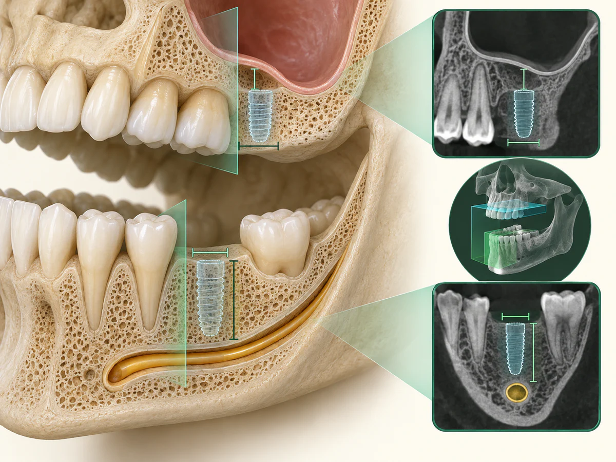
Jacobs (2018) describes CBCT’s role in implant planning. AAOMR (2012) recommends cross-sectional imaging for implant sites; ADA/AAOMR 2026 also distinguishes initial assessment from presurgical planning. These US recommendations are not presented as Moroccan legal requirements.
In practical terms, CBCT helps analyze several key elements of the implant work-up:
- the bone volume available, in both height and thickness, at the planned site;
- the position of the inferior alveolar nerve in the lower jaw, whose protection is critical to the safety of the procedure;
- the location of the maxillary sinuses in the upper jaw, which determines in particular whether a sinus lift is needed;
- the presence of residual roots, lesions, or anatomical obstacles.
This analysis is what makes it possible, before surgery, to know whether the bone is sufficient or whether reconstruction is required. The full implant placement process is described in our dental implant placement steps, and the options when bone is lacking are detailed in Dental Implants When Bone Is Lacking: What Are the Options. The Dürr Dental Cone Beam unit used at our practice is one tool in this planning process; its presence describes a piece of equipment — it does not, in itself, constitute a guarantee of results.
When assessment raises questions about bone volume, our guide helps you understand bone grafting before an implant.
Is cone beam imaging standard before implants?
Cone beam imaging has a well-established place in implant planning, but “standard” does not mean automatic. The US recommendations cited above favor cross-sectional imaging for implant sites, and they still ask that each examination be justified. In practice, the clinician first examines you and reviews your existing images, such as a recent panoramic, then decides whether a CBCT is needed and which field of view suits the site.
A CBCT is most useful when the answer depends on the third dimension: the bone thickness at the planned site, the distance to the nerve in the lower jaw, or the floor of the sinus in the upper jaw. Because the recommendations distinguish the initial assessment from presurgical planning, a CBCT, when it is needed, usually belongs to the planning stage rather than to the very first visit.
The Question of Dose: Justification and Optimization
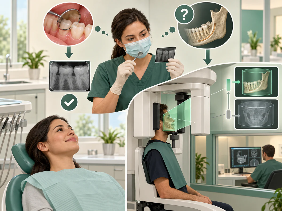
CBCT involves exposure to X-rays. That is why it is never prescribed automatically, but always within the framework of two principles inherited from radiation protection.
The Principle of Justification
Justification means that an examination is performed only if the expected diagnostic benefit outweighs the risk associated with radiation. These fundamental principles — justification and optimization — come from the recommendations of the International Commission on Radiological Protection (ICRP), as outlined notably in the review by Mendonça et al. (2025) on the application of ALARA and ALADA. The European SEDENTEXCT guidelines, published in 2012 by the European Commission (Radiation Protection No. 172), set a clear rule: a CBCT should only be performed after a patient history and clinical examination, and it must be justified for each individual patient, by demonstrating that the benefits outweigh the risks. CBCT is not a routine screening examination.
The Principle of Optimization: ALARA and ALADA
Optimization means delivering the lowest dose compatible with an image that is useful for diagnosis. This is the ALARA principle (As Low As Reasonably Achievable), adapted in dental radiology as ALADA (As Low As Diagnostically Acceptable). As the review by Mendonça et al. (2025) points out, the aim is an image good enough to support a decision — not the most beautiful image possible.
Dose varies with the device, field and settings. It is generally higher than conventional dental X-rays and can be lower than medical CT. Comparisons should concern the examinations and protocols actually considered, not an assumed practice-specific dose.
What CBCT Does Not Provide
An honest presentation of this examination also means stating its limits. CBCT is a tool, not a universal solution.
First, it is not indicated for everything. Many clinical situations can be resolved with a clinical examination and a 2D X-ray, without any need for 3D. Performing a CBCT in the absence of a specific indication would mean exposing the patient to radiation with no expected benefit, which runs counter to the principle of justification.
Second, it is not a guarantee of results. Precise 3D planning improves knowledge of the terrain before a surgical procedure, but it eliminates neither surgical contingencies, nor patient-related risk factors, nor the need for follow-up. CBCT informs the decision; it does not by itself make the treatment a success.
Finally, it has technical limitations of its own. The review by Jacobs et al. (2018) notes that image quality can be degraded by patient movement and by metal artifacts from existing restorations or implants, and that measurement accuracy varies between devices. Results obtained with one CBCT unit therefore do not automatically carry over to another.
What Other Indications Are There Beyond Implantology?
The implant work-up is not the only area where 3D imaging provides a useful complement — always within the framework of a justified indication.
Impacted Teeth
CBCT helps locate an impacted tooth precisely — a tooth that has remained in the bone, such as a wisdom tooth or a canine — and analyze its relationship to neighboring structures. For a lower wisdom tooth, it can clarify how close the root lies to the inferior alveolar nerve when the 2D X-ray leaves room for doubt, which helps assess the risk before extraction. This examination is only justified, however, in cases where 2D imaging is not enough; the extraction process is covered in Wisdom Tooth Extraction: Procedure and Recovery.
Complex Endodontics
The AAE/AAOMR 2025 update, announced in January 2026, replaces the 2015 statement. It recommends selective endodontic use based on clinical findings, existing imaging and patient needs. Field and exposure are adapted to the diagnostic question; CBCT is not routine for every root canal treatment.
In Summary
Cone beam CT (CBCT) is a valuable 3D imaging examination when a specific question goes beyond what a two-dimensional X-ray can show. In the implant work-up, it makes it possible to assess bone volume and locate the inferior alveolar nerve and the sinuses before surgery; it also has a place in analyzing impacted teeth and in certain complex endodontic situations.
But it is neither a routine examination nor a guarantee of results. Its use follows two principles: justification, which reserves the examination for cases where the benefit outweighs the radiation risk, and optimization, which aims for the lowest dose compatible with a reliable diagnosis (ALARA, ALADA). At our practice, the Dürr Dental Cone Beam unit operates within this framework: a tool in the service of a considered decision, prescribed when it is useful — not simply because it is available.
Frequently asked questions
What is the difference between a cone beam CT and a panoramic dental X-ray?
Is a cone beam CT necessary before every implant?
Does a cone beam CT deliver a lot of radiation?
Does a cone beam CT guarantee the success of an implant?
What is a cone beam CT used for besides implants?
Is a cone beam CT suitable for children?
Is a “3D beam” or “3 beam” dental scan the same as a cone beam CT?
Sources
Medical references consulted for this article.
- 1Jacobs et coll. 2018, recommandations sur le CBCT en implantologie, BMC Oral Health 18:88
- 2Tyndall et coll. 2012, AAOMR position statement on radiology in dental implantology with emphasis on CBCT, OOOO (PubMed)
- 3SEDENTEXCT, Radiation Protection N°172: Cone Beam CT for Dental and Maxillofacial Radiology, Evidence-based guidelines, Commission européenne 2012
- 4AAE/AAOMR, Use of Cone-Beam Computed Tomography in Endodontics — 2025 Update
- 5Mendonça et coll. 2025, revue critique des principes ALARA et ALADA en radiologie dentaire
- 6FDA, Dental Cone-beam Computed Tomography (dose, sensibilité des enfants, ALARA)
- 7OMS, Santé bucco-dentaire (aide-mémoire, 17 mars 2025)
- 8Dürr Dental, VistaVox S : système d'imagerie 3D / CBCT dentaire (fiche produit)
- 9AAE/AAOMR, annonce officielle du remplacement de la recommandation de 2015, janvier 2026
- 10ADA/AAOMR, recommandations de sélection des patients pour l'imagerie dentaire, 2026
Contents
Share