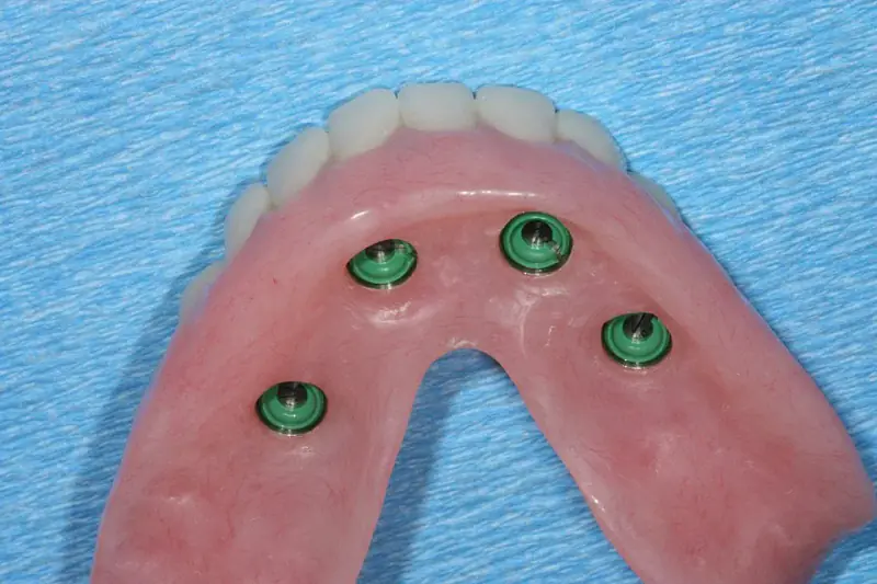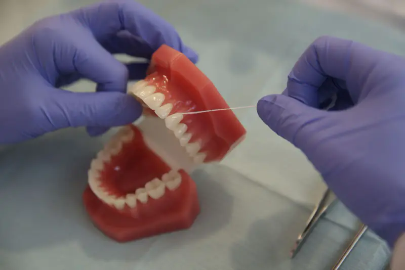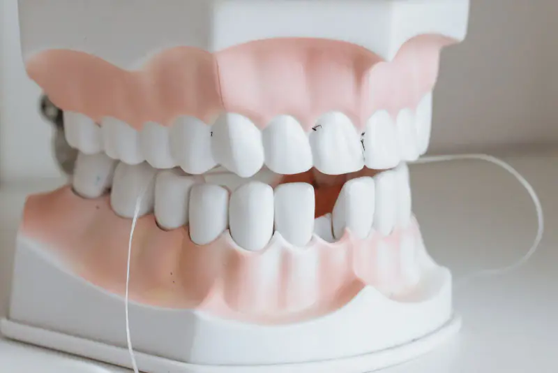
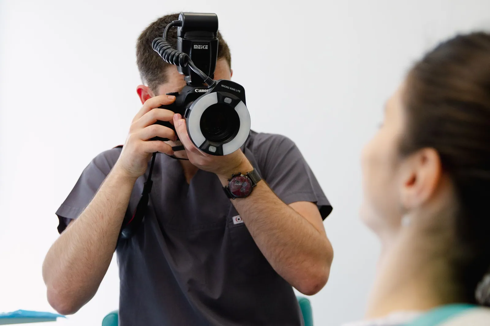
Dental photography means taking precise images of your teeth, your gums and your smile with a camera set up for the job. These aren’t keepsake snapshots: they are a working tool that helps the practitioner examine your mouth more closely, explain what she is seeing, and track how things change over time. At the practice in Kénitra, these images are part of your record, in the same way as consultation notes. This article sets out, simply and honestly, what they are used for, how they are taken, how they are protected, and what they cannot do.
Let’s be clear from the outset: a good photo doesn’t treat anything, and it doesn’t replace an examination. It sheds light on one stage of care; it doesn’t make the decision for you. Its value lies in making visible and shareable a piece of clinical information that would otherwise be hard to put into words.
What dental photography is used for
The uses of dental photography are well described in the professional literature. They fall into a few broad, complementary roles.
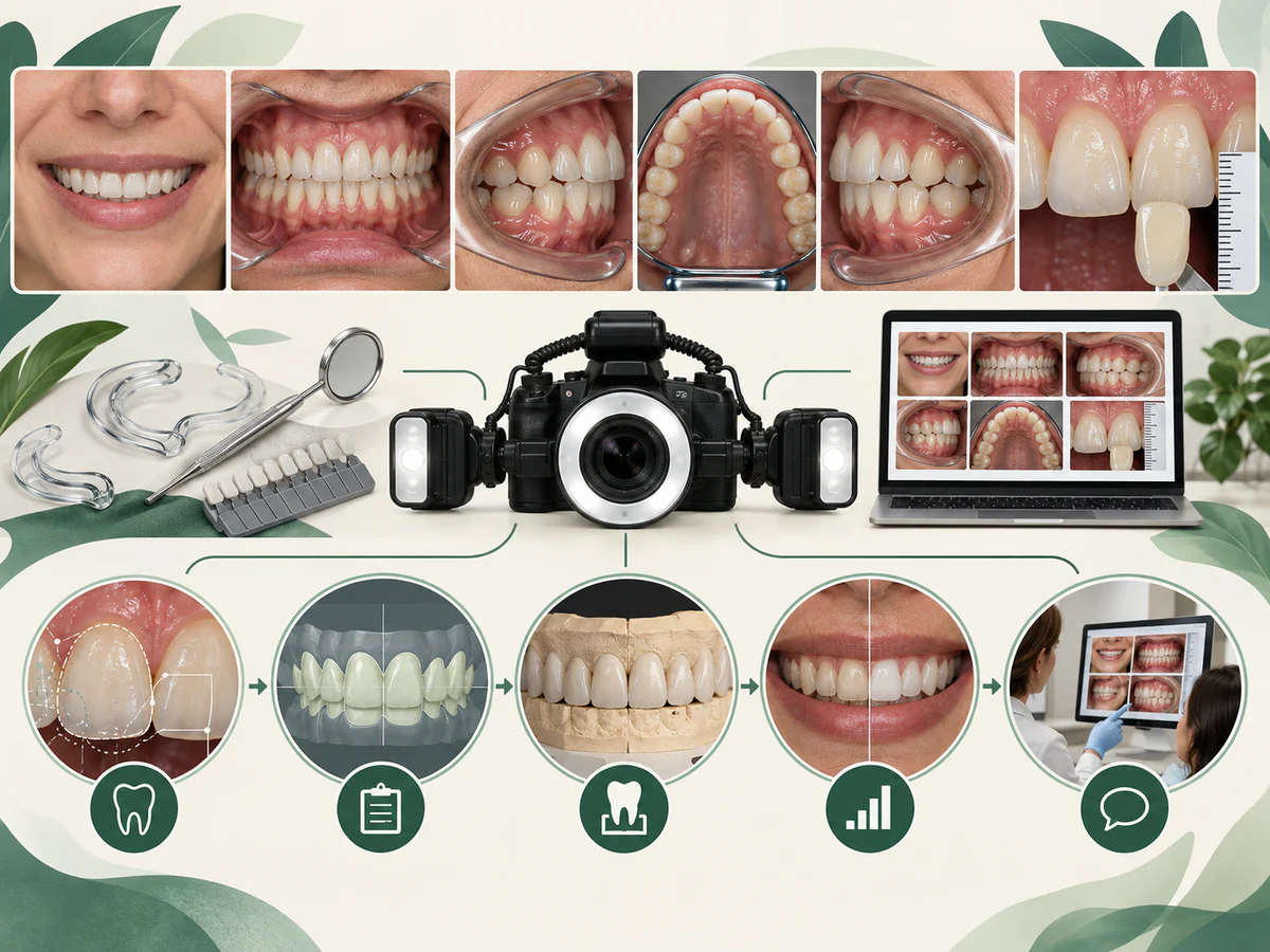
Diagnosis and examination. A good-quality image, enlarged on a screen, reveals details the eye can miss during a quick examination: a crack in the enamel, the beginning of a stain, wear, a gum that is receding slightly. Taking photos also makes it possible to look at areas that are hard to see directly. Photography doesn’t make the diagnosis, but it helps document and refine it.
Communication with you. It is easier to understand a situation when you can see it for yourself rather than picture it from a verbal explanation. Seeing the actual state of your teeth helps you grasp why a treatment matters and take part in the decision. This is a point research regularly underlines: images, and before/after comparisons in particular, strengthen trust and engagement with treatment.
Follow-up over time — before and after. By comparing photos taken several months apart, you can track how a gum, a stain or a restoration is changing. Before a treatment, you see the starting point; afterwards, you compare. It is transparent, and it helps you assess the outcome together — after a cosmetic treatment such as ceramic veneers or teeth whitening, for example.
The link with the dental laboratory. For a crown, a bridge or veneers, color and shape don’t travel well over the phone. Calibrated photos help the dental technician choose the right shade and reproduce the natural nuances of a tooth. Photography then complements the other data in your record, such as the digital impression, to prepare a more faithful restoration.
Aesthetic planning (Digital Smile Design). In some smile projects, design software — grouped under the term “Digital Smile Design” — uses photographs of the face and teeth to simulate and discuss a result before anything is done. The available reviews indicate that this image-based approach most often improves communication, satisfaction and the sense of predictability patients experience. These are tools for discussion and planning: a simulation remains a projection, never a guarantee of the final result.
How it works during the appointment
It is a simple, painless and quick step. The practitioner uses a camera suited to dental photography, often fitted with a ring flash or side flashes that light the inside of the mouth evenly. Small, soft plastic retractors may be placed to hold the lips and cheeks out of the way and open up the view; small mirrors make it possible to photograph the inner surfaces or the tops of the teeth.
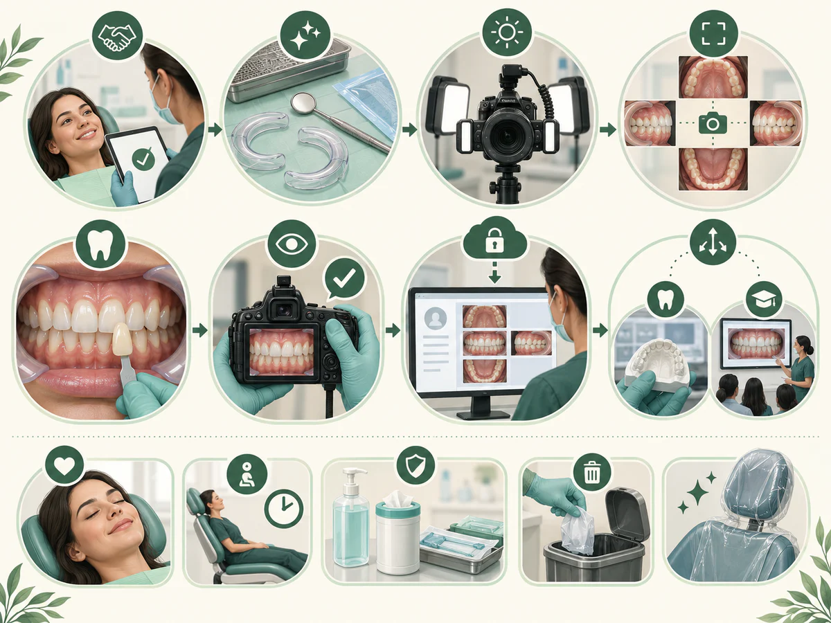
Several angles are recorded depending on what is needed: the face and the smile, the teeth from the front and from the side, the top of the arches, sometimes with the teeth together and then slightly apart. The whole thing usually takes a few minutes. The images are then filed in your digital record and can be displayed on a screen so they can be shown and talked through with you, either on the spot or at a later visit.
In children, these photos can also be used, without any particular constraint, to show parents a specific point or to follow a tooth as it develops — in the spirit described in our article on the child’s first dental visit.
Confidentiality and consent for images
Your dental photographs are health data. They form part of your record and are covered by professional confidentiality, like the rest of your medical information. Several principles govern how they are used, and the professional literature is consistent on this point.
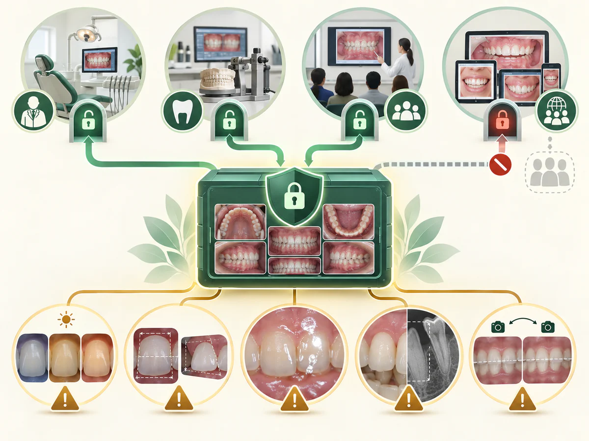
- Consent is specific to each use. Recording photos in your clinical record is one thing; using them for teaching, publication or on social media is quite another. Each of these uses requires your explicit and separate agreement.
- Consent is still required even if the image is anonymized. The fact that you cannot be recognized does not remove the need to ask your agreement before an image is shared publicly.
- Images are stored securely and are shared — with a dental laboratory or a colleague, for instance — only as part of your care and with the appropriate protections.
In practical terms, your photos remain first and foremost a clinical tool serving your follow-up. Any other use is explained to you and requires your authorization; you can refuse without that changing anything at all about the quality of your care.
Its limits
Dental photography is useful, but it is worth knowing its boundaries so as not to credit it with powers it doesn’t have.
A photo is not a diagnosis. An image shows a surface at a given moment. It says nothing about what is happening inside the tooth or beneath the gum, where only a clinical examination and, where appropriate, an X-ray provide the information. An early cavity between two teeth or a bone problem may not be “visible” on a photo. The image documents and illustrates; it replaces neither the examination nor imaging.
Quality and interpretation matter. Lighting, angle or a color setting can change how a tooth looks on a screen. A shade will appear different depending on the light. That is why a photograph should always be read by a professional, alongside the examination, and never in isolation.
A simulation is not a promise. Visual smile projects help you picture the outcome and discuss it, but a simulation shows an intention, not a guaranteed result. The actual result depends on the indication, on the condition of the teeth and gums, on the laboratory work and on follow-up.
An image alone is not a reason to treat. Seeing an area worth watching is useful, but the decision to treat, to monitor or to hold off comes from the overall clinical assessment, not from a single photo, however telling it may be.
In summary
Digital dental photography is a simple, honest tool that makes your care more transparent: you see what the practitioner sees, you understand better, and you decide together. It supports diagnosis, before/after follow-up, dialogue with you and with the dental laboratory, and aesthetic planning. Its images belong to you within your record and are not used beyond your care without your explicit agreement. But a photo is neither a diagnosis nor a guarantee of results: it sheds light on clinical work without ever replacing it. If you have questions about these images or how they are used, do feel free to raise them at your consultation in Kénitra.
Frequently asked questions
What are the photos of my teeth used for?
Is dental photography painful or unpleasant?
Can a photo replace an examination or an X-ray?
What happens to my dental photos, and who can see them?
Does a smile simulation guarantee the final result?
Sources
Medical references consulted for this article.
- 1Ahmad I., Digital dental photography. Part 2: Purposes and uses, British Dental Journal, 2009
- 2Christensen GJ, Digital photography enhances diagnostics, communication, and documentation, JADA (Journal of the American Dental Association), 2011
- 3Jafri Z et al., The Recent Use, Patient Satisfaction, and Advancement in Digital Smile Designing (revue systématique), Cureus, 2024
- 4Digital Smile Design and Patient-Centered Outcomes in Esthetic Restorative Dentistry (revue systématique), Cureus, 2025
- 5Oxholm Gordon J. et al., Considerations for use of dental photography and electronic media in dental education and clinical practice, Journal of the American Dental Association, 2015
- 6Dental photography: data protection and social media, BDJ In Practice (British Dental Journal), 2024
Contents
Share