In short: Digital dental radiography captures X-ray images of the teeth with an electronic receptor instead of film: either a digital sensor or a PSP plate. The image appears on screen within seconds, can be enlarged and archived, and the radiation exposure is generally lower than with traditional film. It is still an X-ray, taken only when it answers a clinical question.
Digital dental radiography refers to all the techniques that replace silver-halide film with an electronic sensor: the image is no longer developed in a chemical bath, it appears directly on a screen a few seconds after being taken. This shift, which has become widespread in dental practices over the past twenty years or so, brings three main benefits: a radiation dose that is generally lower than with the older film, an image that is immediately available and can be manipulated, and digital archiving that makes it easier to follow a patient over time. That said, going digital does not change the nature of the examination: it is still an X-ray, which involves exposure to radiation and is only worthwhile when it answers a specific clinical question.
This article explains, in plain terms, what a digital dental X-ray is, how it differs from conventional film, what the literature tells us about radiation dose and image quality, and — a key point — what this technology does not provide. The aim is not to present digital imaging as a guarantee of absolute precision, but to give honest, nuanced information about an examination that has become routine.
What is a digital dental X-ray?
An X-ray image is always obtained by passing a beam of X-rays through the tissues: bone and teeth, being denser, block more of the radiation and appear light; soft tissues and cavities, which let the radiation through, appear darker. What changes with digital imaging is only the receptor that captures the image, not the underlying physics.
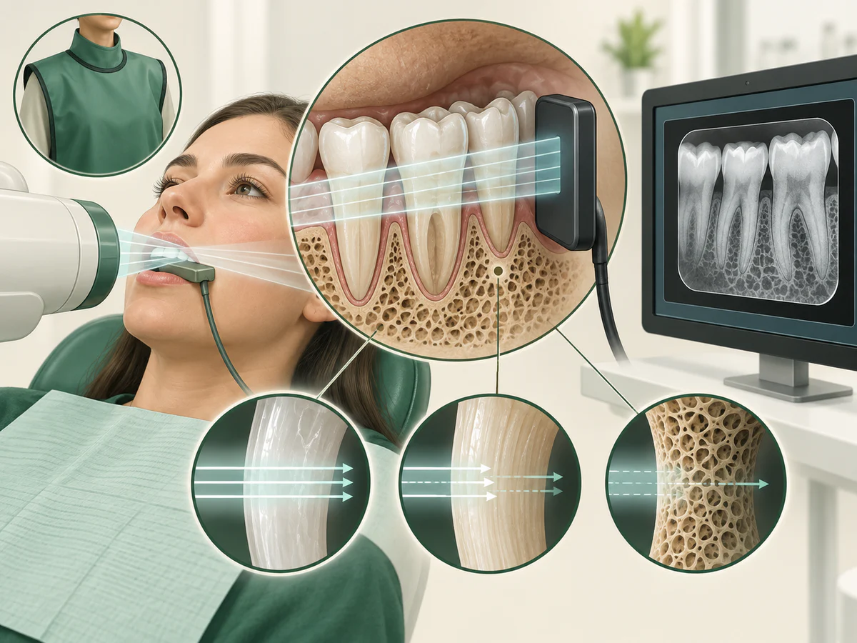
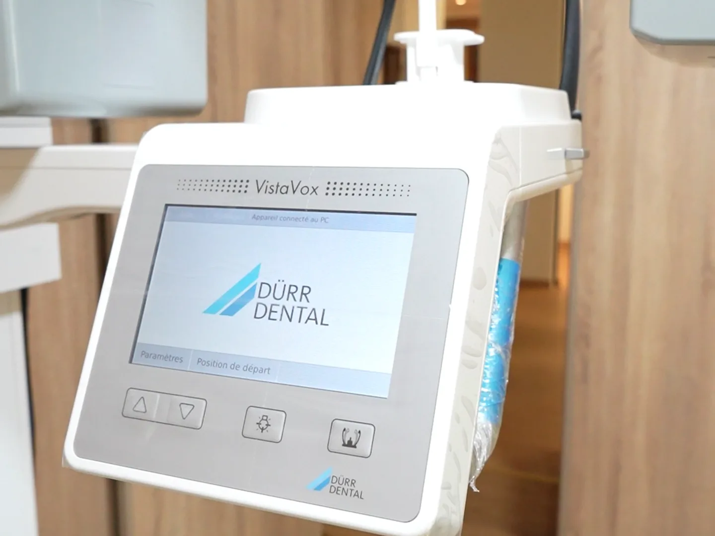
There are two main families of digital receptors. Solid-state sensors (CCD or CMOS technologies) are connected to the computer, usually by a cable, and display the image almost instantly. Photostimulable phosphor (PSP) plates are closer to a flexible film: they are placed in the mouth like a conventional X-ray, then passed through a reader that digitises the image. Both replace silver-halide film and its chemical developing.
These receptors are used for the same examinations as traditional radiography: the periapical X-ray, which shows one or a few teeth in detail, and the digital panoramic X-ray (orthopantomogram), which gives an overview of both arches, the sinuses and the joints. Digital technology also applies to 3D imaging, covered separately in Cone Beam (CBCT): 3D imaging in the practice.
The types of images in digital dental radiography
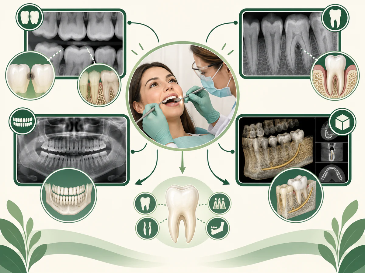
Digital dental radiography uses the same types of images as film did; only the receptor changes. Each type answers a different question:
- Periapical image: one or a few teeth from crown to root tip, with the surrounding bone. It is used to look at a root, an infection at the tip of a root or a root canal treatment.
- Bitewing image: the crowns of the upper and lower back teeth on one image, with the bone level between them. It is the usual image for looking for decay between the teeth.
- Occlusal image: a wider view of the palate or the floor of the mouth, taken with the receptor held between the teeth. It is used less often, for example in young children.
- Panoramic image: both jaws on a single image, taken by a machine that turns around the head.
Your dentist chooses the image that answers the clinical question with the fewest exposures.
Digital or silver-halide film: what actually changes
Going digital mainly changes ease of use and radiation protection, without transforming the way the image is read. Three differences stand out.
First, the image is immediate: there is no developing time and no chemicals, and the X-ray appears on screen within a few seconds. Second, the image can be worked on: it can be enlarged, its contrast adjusted, a distance measured. Third, it is easy to archive and share, which helps compare the situation from one appointment to the next. The table below summarises these differences, without presenting film as “bad”: it served patients well for a long time.
| Criterion | Silver-halide film | Digital radiography |
|---|---|---|
| Obtaining the image | Chemical developing, a few minutes | Displayed on screen, a few seconds |
| Radiation dose | Varies with film speed | Often lower (CCD/CMOS sensors) |
| Image processing | None once developed | Zoom, contrast and measurements possible |
| Archiving and sharing | Physical storage of the film | Digital file, easier comparison |
One point that is often oversimplified deserves qualifying: film was not, in itself, synonymous with a high dose. Fast film, known as “F-speed”, already reduces exposure markedly compared with older, slower films. As the US Food and Drug Administration (FDA) points out in its recommendations on patient selection, using the fastest receptor compatible with diagnosis — F-speed film or digital — is one of the pillars of good practice. Digital imaging therefore brings a real dose reduction, but one that belongs within a wider set of measures rather than standing as an isolated advantage.
For a dental practice, the change also has a practical side: digital equipment involves start-up costs (sensors or plates, reading software, screens), whereas traditional dental films need a regular supply of film and developing chemicals. For you as a patient, these are background details. What matters is an image that answers the clinical question at the lowest dose compatible with a useful reading.
Radiation dose: what does the literature say?
This is probably the most frequently cited argument, and it deserves to be qualified rather than overstated. Several studies confirm that digital imaging does, in practice, allow a reduction in dose compared with conventional film. A study carried out in general dental practices by Anissi and Geibel (2014), published in the journal Rofo, compared digital systems with film and observed a significant reduction in dose with sensors, despite a slight increase in the number of images taken. The systematic review by Ardila et al. (2024), published in Dentistry Journal, concludes for its part that digital sensors, both wired and wireless, deliver doses well below those of conventional radiography, while producing high-quality images.
It is worth putting these figures in perspective. The review of doses in dental imaging by Lee and Badal (2021), published in Radiology Research and Practice, reports an average effective dose of about 1.3 microsieverts (µSv) for an intraoral X-ray and about 18 µSv for a panoramic. For comparison, the International Atomic Energy Agency (IAEA) points out that these levels are low relative to the natural background radiation we receive from our environment every day. This does not mean that an examination is “radiation-free” — none is — but that the dose from a digital dental X-ray is, in absolute terms, modest when the examination is justified.
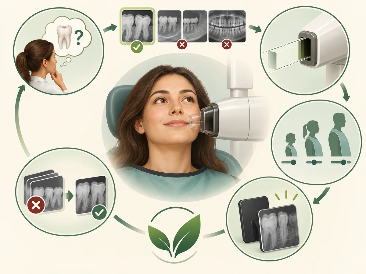
Two principles govern that dose, whatever the receptor. The principle of justification: an examination is performed only if the expected diagnostic benefit outweighs the risk associated with the radiation. The principle of optimisation, summed up by the acronym ALARA (“As Low As Reasonably Achievable”): the aim is the lowest dose compatible with a useful image. Digital imaging serves these principles; it does not replace them. Radiation safety therefore depends on when and how an X-ray is taken, not only on the type of receptor.
What affects the radiation dose of a digital dental X-ray?
The radiation dose of a digital dental X-ray depends less on the word “digital” than on how the image is taken. A few factors make the difference:
- The type of image. A single intraoral image delivers less than a panoramic, as the figures above show, and a 3D Cone Beam delivers more again.
- The receptor and the settings. The reduction is most often reported with solid-state sensors. The exposure is also adjusted to the area and to the patient, with lower settings for a child, and the beam is narrowed to the zone being examined.
- The number of images. Because digital images are quick to take, it can be tempting to take more: the study by Anissi and Geibel cited above observed a slight increase in their number. Careful positioning also avoids having to retake an image.
In other words, the lowest dose comes from a justified examination, taken once, with settings suited to you.
Image quality and diagnostic reliability
A lower dose would be of little value if it came at the cost of a poorer reading. The available data are reassuring on this point, without making digital imaging a “superior” examination in every situation.
For the detection of cavities between the teeth (approximal caries), the comparison between digital and film has been the subject of a great deal of research. The review by Abesi et al. (2012), published in the Iranian Journal of Radiology, concludes that the diagnostic accuracy of digital images is comparable to that of silver-halide film for non-cavitated approximal caries. In other words, digital performs at least as well as film, but does not radically transform the ability to see an early lesion.
This is an important point to keep in mind: no X-ray, digital or otherwise, detects every early cavity. Studies show that sensitivity remains limited for enamel lesions, which can go unnoticed on the image. The X-ray complements the clinical examination; it does not replace it. The diagnosis and management of a cavity are set out in detail in Adult tooth decay: from diagnosis to treatment; there, the X-ray is only one tool among others.
The adjustment options offered by a digital image (contrast, magnification) help the practitioner, but they do not create information that is absent from the image. Image processing does not invent a detail the X-rays did not record: it only makes what was captured easier to read. Sensors are often described as high-resolution, but even sharp images of teeth are only useful when the receptor is correctly positioned and the image is read with care.
Digital radiography in dentistry: bone loss and gum disease
Dental X-rays are not only used to look for cavities. They also show the bone that holds the teeth in place. In periodontal disease (periodontitis), the form of gum disease that reaches the deeper tissues, this bone can be gradually lost, and the X-ray shows how far the bone level has dropped around each tooth. Compared to traditional films, digital images make this follow-up easier: the bone level can be enlarged on screen and set side by side with earlier images to see whether it has remained stable.
An X-ray does not replace the examination of the gums, however. Gum inflammation that has not reached the bone (gingivitis) does not show on an X-ray, and measuring the pockets around the teeth with a fine probe remains essential. The X-ray shows the bone, not the gums themselves. The difference between the two conditions is explained in Periodontitis or gingivitis: understanding the difference.
What digital radiography does not provide
An honest account also means setting out the limits of this technology, so that nothing more is expected of it than it can offer.
Digital imaging does not do away with radiation. It often lowers the dose, but an X-ray remains an X-ray: it is justified by a clinical question, and not carried out routinely “just to take stock”. The 2026 recommendations of the American Dental Association (ADA) and the American Academy of Oral and Maxillofacial Radiology (AAOMR), published in Oral Surgery, Oral Medicine, Oral Pathology and Oral Radiology, state that every radiographic examination — digital included — should be prescribed after a clinical examination and an assessment of individual risk, and not at fixed intervals for everyone.
Digital imaging does not in itself guarantee a better diagnosis. The quality of an X-ray depends first and foremost on positioning, settings and interpretation, far more than on the mere fact that it is digital. Poor framing gives an image of little use, whether on film or on a sensor.
Digital imaging replaces neither the clinical examination nor, when it is indicated, 3D imaging. A 2D X-ray, even a digital one, superimposes structures and does not measure the actual thickness of the bone: for certain questions, particularly implant planning, it is Cone Beam (CBCT) that supplies the missing volume, within the framework of a justified indication. Finally, perceived comfort varies: the review by Ardila et al. (2024) notes that rigid sensors are not systematically more comfortable than film for the patient, which puts the idea of “total” progress into perspective.
In practice, for the patient
From the patient’s point of view, the experience of a digital X-ray remains close to that of a conventional one: a sensor or a plate placed in the mouth for intraoral images, or a machine that rotates around the head for the panoramic. The main visible difference is how quickly the image appears on the screen, which the practitioner can then talk through straight away. Because X-rays show the bone as well as the teeth, digital radiographs kept in your file can be compared at later visits and used, alongside the clinical examination, when your dentist explains the possible treatment plans.
The usual radiation protection measures continue to apply: keeping the number of images to the strict minimum, and appropriate protection depending on the situation, particularly in children, who are more sensitive to radiation, and in pregnant women, for whom any prescription is weighed with particular care. These imaging examinations help inform an overall assessment, whether that is follow-up of treatment, a periodontal assessment or preparation for a dental implant in Kénitra. Imaging is a means in the service of a decision, never an end in itself.
In summary
Digital dental radiography replaces silver-halide film with an electronic sensor: the image appears on screen immediately, can be worked on and archived easily, and the radiation dose is generally lower than with the older film, as the work of Anissi and Geibel (2014) and the review by Ardila et al. (2024) confirm. Its diagnostic accuracy is comparable to that of film for the detection of cavities (Abesi et al., 2012), and the doses involved remain modest when the examination is justified (Lee and Badal, 2021; IAEA). But digital imaging does not do away with radiation, does not on its own guarantee a better diagnosis, and replaces neither the clinical examination nor, where relevant, 3D imaging. As the 2026 ADA and AAOMR recommendations stress, an X-ray — digital included — is justified only by a specific clinical question, in keeping with the principles of justification and ALARA.
Frequently asked questions
What is the difference between a digital dental X-ray and conventional film?
Does a digital X-ray deliver less radiation than film?
What is the radiation dose of a digital dental X-ray?
Does a digital X-ray see cavities better?
Does a digital panoramic X-ray replace other examinations?
Is a dental X-ray dangerous during pregnancy or in children?
Can my dentist show me my digital X-rays?
What is the difference between digital sensors and PSP plates?
Sources
Medical references consulted for this article.
- 1Lee & Badal 2021, A Review of Doses for Dental Imaging in 2010-2020 and Development of a Web Dose Calculator, Radiology Research and Practice (PMC)
- 2Ardila et coll. 2024, Image Quality, Radiation Dose, and Patient Comfort Associated with Wireless Sensors in Digital Radiography: A Systematic Review, Dentistry Journal (PMC)
- 3Anissi & Geibel 2014, Intraoral radiology in general dental practices - a comparison of digital and film-based X-ray systems with regard to radiation protection and dose reduction, Rofo (PubMed)
- 4Abesi et coll. 2012, Diagnostic Accuracy of Digital and Conventional Radiography in the Detection of Non-Cavitated Approximal Dental Caries, Iranian Journal of Radiology (PMC)
- 5ADA & AAOMR (Benavides et coll.) 2026, Patient selection for dental radiography and cone-beam computed tomography: clinical recommendations, Oral Surgery Oral Medicine Oral Pathology and Oral Radiology (PubMed)
- 6FDA, The Selection of Patients for Dental Radiographic Examinations (ALARA, récepteur le plus rapide, sensibilité des enfants)
- 7AIEA (IAEA), Radiation doses in dental radiology - FAQs for health professionals
- 8American Dental Association, Radiographic Imaging et ALARA (Oral Health Topics)
Contents
Share