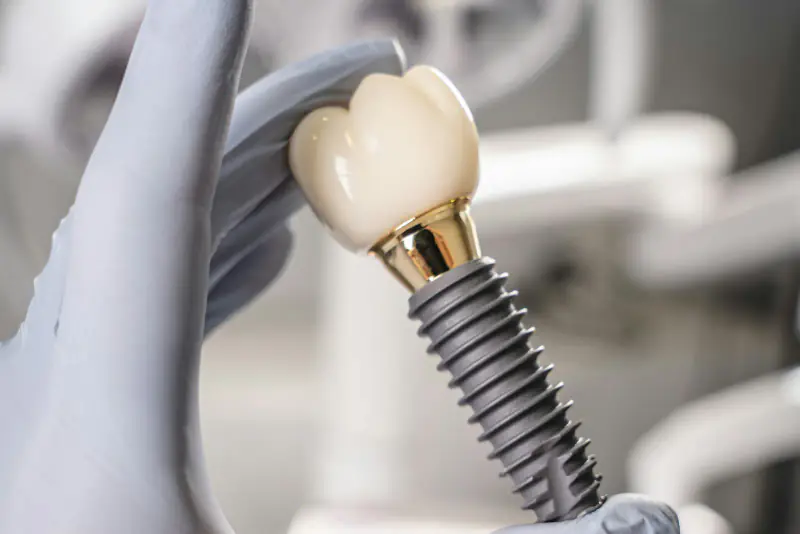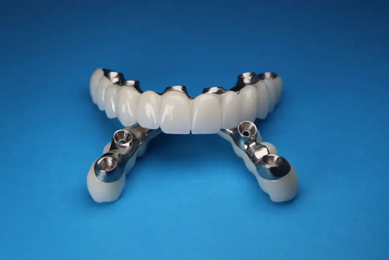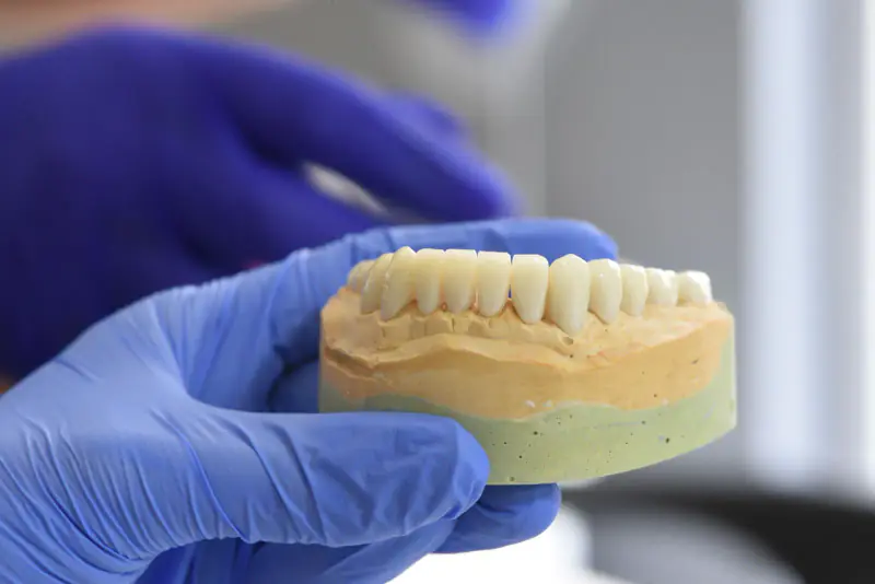
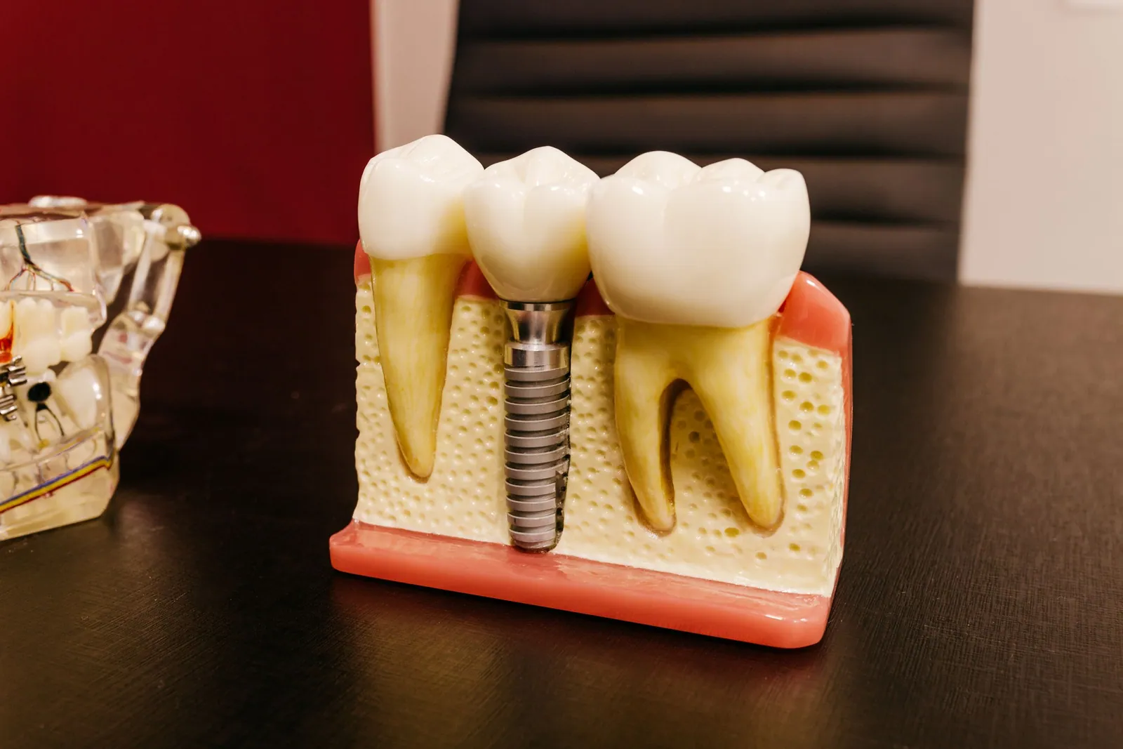
A bone graft before an implant involves rebuilding the volume of jawbone when it has become insufficient to anchor an artificial root stably. It is proposed when the examination, in particular the Cone Beam scan, shows a bone height or thickness below what the implant requires. It is neither routine nor trivial: it is a surgical procedure in its own right, with precise indications, several techniques and after-effects to anticipate. This article explains why bone is lacking, how it is rebuilt, what can realistically be expected from healing, and which factors influence the outcome.
Why does the jawbone shrink after a tooth is lost?
As long as a tooth is in place, its root stimulates the surrounding bone and maintains it. When the tooth is extracted or lost, that stimulation disappears and the alveolar bone, the bone that formed the socket, resorbs.
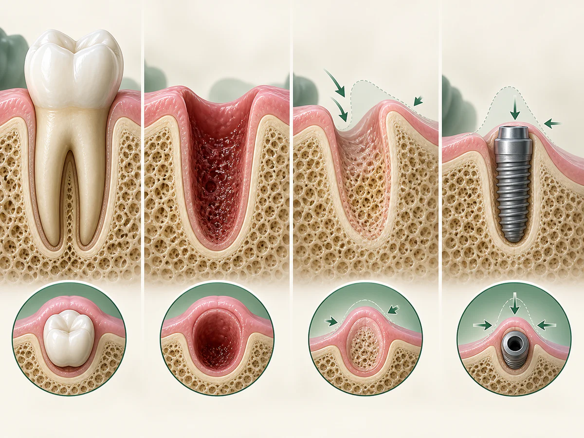
This loss is measurable. According to the systematic review by Tan and colleagues, published in Clinical Oral Implants Research in 2012, at six months an average horizontal loss of about 3.79 mm is observed, that is 29 to 63% of the thickness, along with an average vertical loss of about 1.2 mm on the buccal side, that is 11 to 22% of the height. Most of it occurs during the first three to six months following the extraction.
Remodelling varies between sites; elapsed time alone cannot determine whether grafting is needed. See the options for implants when bone is insufficient.
When is a bone graft indicated before an implant?
Grafting is discussed when the bone volume does not allow an implant to be placed in a correct position with sufficient initial stability. The following situations are common.
- A ridge too thin to fully surround the implant.
- Insufficient bone height, particularly in the posterior maxilla where the sinus descends.
- A localized defect following an old extraction, a cyst or an infection.
- Bone loss related to advanced periodontal disease.
In the posterior maxilla, a lack of height beneath the sinus calls for a specific technique, sinus floor augmentation, covered in our dedicated article on sinus augmentation, or sinus lift.
Grafting is not the only possible answer. In some cases, shorter or tilted implants, a different site, or a different prosthesis may be chosen. This nuance matters: the Cochrane review by Esposito and colleagues (2009) points out that vertical augmentations are associated with more complications than the placement of short implants. The choice is made case by case, after an assessment.
Grafting, GBR, block graft, gum graft: what does each word mean?
These words describe different procedures. Here they are in plain terms, with the labels used on the Fees page.
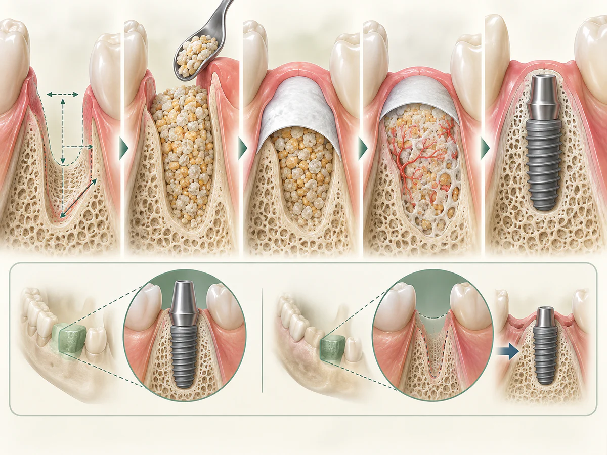
- Bone grafting (simple graft). A small hollow in the bone, for example where a tooth has been removed, is filled with granules of graft material. When this is done on the day of an extraction to limit bone shrinkage, it is called socket preservation: whether to fill the socket at the time of extraction is explained separately.
- Guided bone regeneration (GBR). The material is covered with a membrane, a thin barrier that protects the space while bone forms. It is used to regain width, sometimes height, on a ridge that has become thin.
- Block graft. A single piece of bone is fixed against the ridge with small titanium screws. It is taken from you, for example from the chin or near the lower wisdom tooth, or it comes from a bone bank. It is used for larger defects. According to the French Society of Oral Surgery, implants are placed on average four months later, and the screws are removed at that time. Because it is placed against the ridge, it belongs to the so-called onlay (apposition) grafts.
- Sinus lift (sinus augmentation). A graft placed under the sinus, at the back of the upper jaw, when height is lacking there.
- Gum graft. This is not a bone graft. Gum tissue, most often taken from the palate, is added to cover an exposed root or to thicken thin gum. It belongs to gum treatment, described in the article on gum recession.
From assessment to the decision: what is checked
01. Assess the situation and the replacement plan
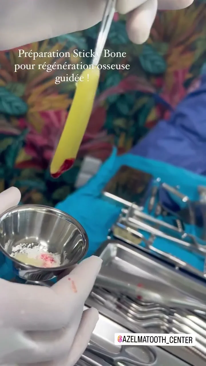
Assessment combines examination, medical history and the replacement plan. Imaging, when indicated, adds information. Values visible on a photograph are not universal thresholds for self-assessment.
02. Compare the options
Ask why reconstruction is proposed and which alternatives were discussed. The plan should say whether grafting is envisaged before or during implant placement. Leeds Teaching Hospitals describes these possible pathways, without allowing a patient’s treatment to be chosen remotely.
03. Understand the material and any device
A graft, membrane and fixation device have different roles. Ask what will be used, its origin, purpose and whether removal may be necessary. Appearance in an operative photograph cannot reliably identify a material or its function.
04. Arrange healing and follow-up
Ask for written instructions, a contact for difficulties and the date for reassessment. A photograph cannot establish completion of treatment; appropriate clinical follow-up is needed.
Read about Cone Beam images and the dental implant placement steps.
To prepare for your consultation, see our information on dental bone grafting in Kénitra.
What are the types of dental bone graft material?
Grafts are generally divided into four groups by origin. They may be used alone or together. Properties, indications and risks differ; no material is best for every situation.
| Graft type | Origin | Key points |
|---|---|---|
| Autogenous | Bone harvested from the patient | Biological reference standard; requires a donor site |
| Allogeneic | Processed and safety-treated human bone | Avoids a second harvest site |
| Xenogeneic | Processed bone of animal origin | Acts as a scaffold; slow resorption |
| Alloplastic | Synthetic material, for example tricalcium phosphate | Entirely manufactured; no donor |
The autograft, the biological reference
Autogenous bone comes from the patient and can provide living cells. Harvesting creates another operative site with its own recovery and risks. Its benefits must be weighed against other options for the defect.
Allografts, xenografts and synthetic materials
Allogeneic, xenogeneic and alloplastic grafts avoid this second site. They mainly serve as a support, a scaffold that the body gradually colonizes with its own bone. Their resorption rate and their behavior differ from one material to another, which guides the choice according to the defect to be treated.
One point deserves clarification: the CE marking of a biomaterial is a regulatory conformity for placing it on the market, not proof of clinical efficacy nor a guarantee of results. The predictability of augmentation techniques rests on consensus data, not on a commercial label.
What is guided bone regeneration and the role of the membrane?
Guided bone regeneration (GBR) uses a barrier, usually a membrane, to protect the space intended for bone formation from soft-tissue ingrowth. It may be combined with a graft material.
Membrane choice, stabilisation and any removal depend on the defect and technique. A membrane alone does not guarantee regeneration.
Not all bone augmentation is GBR. The ITI 2008 passage mentioning coverage with or without a membrane concerns onlay grafting; it does not remove the barrier principle underlying GBR.
How does a dental bone graft procedure work?
The procedure is carried out under local anesthesia, in sterile conditions, as described by the ITI consensus. The general sequence includes accessing the area, preparing the recipient site, placing the graft and possibly a membrane, then closing.
Two approaches exist as to when the implant is placed.
- Simultaneous approach, one stage. The implant is placed at the same time as the graft, when the residual bone allows sufficient primary stability to be achieved.
- Staged approach, two stages. The graft is performed first, healing is allowed to take place, then the implant is placed. The ITI consensus favors delayed placement for onlay grafts.
The full course of the implant phase, once the bone has been rebuilt, is described in our article on dental implants in Kénitra.
The day of the procedure, in practice
Under local anaesthesia, you stay awake. A few injections numb the area: the aim is that you feel no pain. You may feel pressure or vibration and hear the instruments. If a sensation becomes painful, say so, for example by raising your hand: the anaesthesia can be topped up.
The session usually follows this order:
- The plan, your medicines and your questions are reviewed with you.
- The local anaesthetic is given, then there is a short wait for it to work.
- The gum is opened to reach the bone.
- The graft is placed, with a membrane or small screws if the technique requires them.
- The gum is closed with stitches, then you go home.
For a graft using a small amount of bone taken from the jaw, the patient information from Guy’s and St Thomas’ hospital (NHS) usually allows one hour to one and a half hours, with the stitches removed about a week later. The actual time depends on the extent; for a sinus lift, see the article on sinus lifts.
If you feel very apprehensive, mention it before the appointment. Ways to help an anxious person are presented in the article on conscious sedation and fear of the dentist.
Choukroun’s PRF as an adjunct
PRF, platelet-rich fibrin following the Choukroun protocol, is an autologous concentrate obtained from a blood sample taken from the patient, then centrifuged. It releases factors that support healing and it can be combined with the graft.
Alrayyes and Al-Jasser (2022) reviewed socket preservation after extraction, not every pre-implant graft. PRF should not be presented as routinely replacing a biomaterial or guaranteeing regeneration. Results depend on the indication and protocol.
How long does healing take after a bone graft?
There is no single timeframe that applies to everyone. Healing depends on the type of graft, the site, the volume rebuilt and the approach chosen.
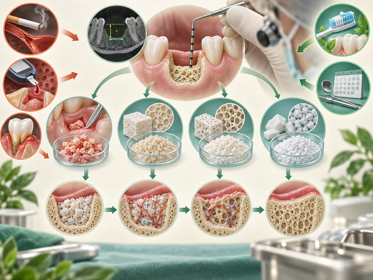
Healing often takes several months depending on technique and clinical assessment. Simultaneous placement may be possible when residual bone provides sufficient initial stability. Follow-up determines timing; older study ranges are not a universal rule.
Presenting a fixed timeframe for every situation would be inaccurate. The best estimate is the one given for your own case, after reading the Cone Beam scan and choosing the technique.
From the first appointment to the final tooth: the timeline with a graft
Each stage has its own timing. Added together, they give an order of magnitude, not a date. Here is the usual path when the graft is done before the implant, in two stages.
| Stage | What happens | Order of magnitude |
|---|---|---|
| Assessment | Examination, imaging if indicated, treatment plan | Before any decision |
| Graft | The procedure described above, then removal of the stitches | One session, then a check-up |
| Graft healing | Bone forms within the material | Often 3 to 6 months (Guy’s and St Thomas’, NHS) |
| Implant placement | Sometimes with a small additional bone graft | One session, then a check-up |
| Implant integration | The bone bonds to the implant | 3 months on average (UFSBD) |
| Final tooth | Impression, try-in, fitting of the crown | Depends on the planned prosthesis |
From the graft to the final tooth, you should therefore often allow around six to ten months, and at least one appointment per stage, plus check-ups. The path is shorter when the graft and the implant are done on the same day, and longer after a sinus lift. Without a graft, it follows the dental implant placement steps.
During healing, nothing should press on the grafted area, not even a removable appliance, for the period you are given. Solutions to avoid a visible gap are described in what to wear while waiting for the implant.
What can be expected in terms of reliability?
No practice-specific success rate can be stated without documented local data and an explicit method. Published results depend on technique, defect and follow-up; they are not an individual guarantee.
What helps a graft to take
A graft takes when your bone gradually grows into the material. Several conditions contribute, and some depend on you.
- A graft that does not move. That is the role of the screws for a block and of the membrane for GBR.
- Gum that stays closed over it. If the wound opens, the graft is exposed and can become infected or be lost.
- Nothing pressing on the area, such as a removable appliance put back too early.
- No smoking. The risk of failure is higher in smokers.
- A clean mouth, healthy gums and well-controlled diabetes, covered below.
Once the implant is in place, the rebuilt bone is protected as around any implant: see what makes an implant last longer and implant follow-up.
What if the graft does not take?
Sometimes a graft does not give all the volume hoped for, or part of it is lost, for example after an infection. The patient information from Guy’s and St Thomas’ puts it simply: sometimes the bone obtained is not enough to place the implant.
This is not a dead end. The check-up, with a new image if needed, measures what has been obtained. Depending on the result, the implant can be placed as planned, bone can be added when the implant is placed, the area can be left to heal before a new reconstruction, or a solution that needs less bone can be chosen.
What are the risk factors and contraindications?
A bone graft is not a procedure without after-effects. Nuance is part of honest information.
Post-operative discomfort is real
Recovery depends on the extent of reconstruction and any bone harvesting. Pain, swelling and healing complications are possible. The clinician will explain procedure-specific risks, aftercare and signs requiring prompt contact.
Smoking
Smoking should be discussed before implant surgery. Chen and Mustapha found associations with more implant failures; their studies do not provide a single failure percentage for your bone graft. Preparation and follow-up are individualised.
A graft can take in someone who smokes, but the risk of failure is higher. Before a procedure, the French stop-smoking service Tabac Info Service says the ideal is to have stopped six to eight weeks beforehand. After the graft, do not start again during healing, and ideally not at all. Shisha counts too: the water does not filter its smoke, which remains highly toxic. If you smoke cannabis, say so as well, so that the instructions take it into account. The article on the effects of tobacco on the mouth explains what the mouth recovers after stopping.
Diabetes
Diabetes does not automatically rule out implants. Disease control, other risks and follow-up require individual assessment; the diagnosis alone cannot predict a person’s outcome. Smoking cessation should be discussed. Insufficient bone calls for comparison of options, not automatically a graft. Wagner 2022.
Pregnancy, blood pressure, blood thinners, age: what to mention
- Pregnancy. A graft before an implant is planned surgery, not an emergency: it is generally scheduled after the birth. Care that cannot wait, such as pain or infection, remains possible at any stage of pregnancy, as the American Dental Association points out. See also dental care during pregnancy.
- Blood pressure. Mention high blood pressure and its treatment. Very high blood pressure on the planned day leads to postponing planned surgery and calls for your doctor’s advice.
- Anticoagulants and antiplatelet drugs, the medicines that thin the blood, aspirin included. Never stop them on your own. According to the Scottish SDCEP guidance (2022), most are continued during dental surgery, with local measures against bleeding; for some, an adjustment is decided, with the prescribing doctor if needed.
- Bone medicines. Mention any treatment for osteoporosis, such as alendronic acid: the NHS states that there is then a very small chance of damage to the jawbone when having dental implants or a tooth out.
- Age. Age alone does not close the door. A 2025 review concludes that implants remain a reliable option for older people, including those over 75. What matters is your general health, your treatments and how you heal.
Other factors
Oral hygiene, the condition of the gums and the control of any periodontal disease also shape the outcome. Active periodontitis must be treated before any surgery; to tell simple inflammation from a deeper condition, see our article on the difference between periodontitis and gingivitis. After placement, monitoring remains important, as explained in our article on the signs of implant failure and peri-implantitis.
The equipment used to plan and perform the graft
Imaging supports planning but does not decide grafting on its own. Ask which examinations, materials and instruments are planned, why they are useful and what their limits are. Ultrasound instruments do not eliminate anatomical risks; any PRF use should be explained for your indication.
In summary
A bone graft before an implant answers a real lack of bone, documented by the examination. It relies on four families of graft material, sometimes combined with guided bone regeneration and a membrane when this is indicated. Healing generally takes several months, within a range that depends on the case. The benefits are established, but the after-effects and the risk factors, particularly smoking, must be taken into account. Honest information is worth more than a promise: the right decision is built after a precise assessment and a discussion of the options.
After-effects vary with the technique and extent of grafting. Pain, swelling, wound opening, infection or altered sensation can occur. Contact the treating team for worsening pain or swelling, fever, discharge or persistent bleeding. Difficulty breathing or swallowing requires immediate emergency assessment: do not wait for a WhatsApp reply. NHS emergency signs.
Frequently asked questions
Is a dental bone graft painful?
How long do you have to wait between the graft and implant placement?
Can an implant be placed without a graft when there is not enough bone?
Does smoking prevent a bone graft from succeeding?
Does PRF replace the graft material?
Can rebuilt bone shrink again over the years?
Sources
Medical references consulted for this article.
- 1ITI, 4th Consensus Conference, Bone augmentation procedures in extended alveolar ridge defects (2008)
- 2Cochrane, Esposito et al., Horizontal and vertical bone augmentation for dental implants, CD003607 (2009)
- 3Tan et al., Post-extraction alveolar dimensional changes, Clinical Oral Implants Research (2012), PMID 22211303
- 4Chen et al., Risk factors for implant failure, PLOS One (2013), PMC3733795
- 5Mustapha, Salame, Chrcanovic, Smoking and Dental Implants, Medicina (2021), PMID 35056347
- 6Alrayyes & Al-Jasser, PRF in Socket Preservation, Tissue Engineering and Regenerative Medicine (2022), PMID 35334092
- 7Wagner et coll. 2022, revue systématique sur le diabète et les implants dentaires
- 8Al Ansari et coll. 2022, revue systématique et méta-analyse sur diabète et implants dentaires
- 9Société Française de Chirurgie Orale, Information médicale : Greffes (fiche patient)
- 10Guy's and St Thomas' NHS Foundation Trust, Bone grafting for dental implants (information patient, revue décembre 2024)
- 11Guy's and St Thomas' NHS Foundation Trust, Minor bone grafting (information patient, revue décembre 2024)
- 12UFSBD, Fiche patient « Un implant pour remplacer une dent », 2016
- 13Tabac Info Service, Cicatrisation : quand arrêter avant une intervention
- 14Tabac Info Service, Risque de la chicha
- 15American Dental Association, Oral Health Topics : Pregnancy (mise à jour 2025)
- 16American Dental Association, Oral Health Topics : Hypertension (2022)
- 17SDCEP, Management of Dental Patients Taking Anticoagulants or Antiplatelet Drugs, 2e édition (2022)
- 18NHS, Common questions about alendronic acid
- 19Abou-Ayash et coll., Implant survival in patients aged 65 to 75 compared to older cohorts, Clinical Oral Implants Research (2025)
- 20Elnayef et coll., The Fate of Lateral Ridge Augmentation, revue systématique, Int J Oral Maxillofac Implants (2018), PMID 29763500
Contents
Share