A dental implant in trouble usually gives warning signs before it gives way. The symptoms to watch for are, first, gum bleeding when brushing or on contact, then local redness or swelling, sometimes a discharge of pus, discomfort or pain, and, later on, mobility of the implant. The key clinical distinction, established by the World Workshop 2017 consensus, sets peri-implant mucositis, a reversible inflammation with no bone loss, apart from peri-implantitis, which involves progressive loss of the supporting bone and is far more difficult to correct. Recognizing the first stage and getting it checked early changes the outlook.
This article describes these signs one by one, explains the factors that raise the risk, and details the maintenance that keeps an implant functional over time. The aim is not to alarm. It is to replace false reassurance with useful vigilance.
Can an implant fail? What the 2017 classification says
Contrary to a widespread idea, a titanium implant is not “rejected” the way an organ graft would be. The dominant biological failure mechanism is different: it is a disease associated with bacterial biofilm, which causes inflammation of the tissues around the implant and then, in its advanced form, destruction of the bone that holds it in place.
The World Workshop 2017, organized jointly by the American and European periodontology societies, introduced for the first time formal clinical definitions for three distinct situations: peri-implant health, peri-implant mucositis and peri-implantitis. This is the reference classification today (Berglundh et al., 2017 consensus). Understanding these three states helps you make sense of what you are feeling.
These conditions are not marginal. The systematic review by Derks and Tomasi (2015) estimates the mean prevalence of peri-implant mucositis at around 43 percent of implant patients, and that of peri-implantitis at around 22 percent, with wide variation depending on the definitions used. In other words, implants are a generally predictable treatment, but one that carries a real biological risk. No surgical procedure is “risk-free,” and it is precisely that honesty that underpins good monitoring.
Peri-implant mucositis or peri-implantitis: what is the difference?
This is the most important distinction in the whole article, because it determines what can still be reversed.
Peri-implant mucositis: a reversible inflammation
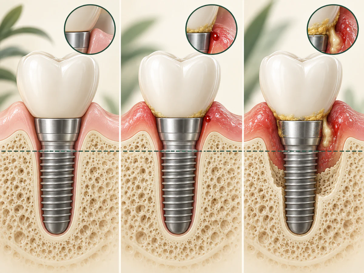
Mucositis is an inflammation of the soft tissues around the implant, without loss of the supporting bone. According to the consensus by Berglundh et al. (2017), its main clinical sign is bleeding on gentle probing; redness, slight swelling and sometimes a discharge may also be observed. The causal factor is plaque accumulation, and there is solid evidence that mucositis is treatable and reversible through rigorous biofilm control. The EFP S3 guideline (Herrera et al., 2023) confirms that mucositis can be managed successfully through careful control of plaque around the implant.
This, then, is the stage at which to act. A gum that bleeds around an implant is never “normal” or “nothing to worry about.”
Peri-implantitis: when the bone is affected
Peri-implantitis is defined as an inflammation of the peri-implant tissues associated with progressive loss of the supporting bone (Berglundh et al. 2017; Schwarz et al. 2018). On top of the inflammatory signs (bleeding on probing and/or suppuration, increased probing depth, sometimes gum recession) comes bone loss visible on radiographs. This bone loss does not repair itself spontaneously, and managing it requires intervention, often surgical at advanced stages.
One point deserves attention: according to Schwarz et al. (2018), peri-implantitis can begin early in follow-up and progress in a non-linear, accelerating pattern, with lesions that are sometimes more extensive than those of periodontitis. This rapid course explains why regular radiographic and clinical checks make full sense.
Finally, mucositis precedes peri-implantitis. The 2017 consensus and the EFP S3 guideline stress that untreated mucositis can progress to peri-implantitis, particularly in the absence of regular maintenance. That is the clinical reason why an early sign should not be ignored.
| Feature | Peri-implant mucositis | Peri-implantitis |
|---|---|---|
| Tissues affected | Gum only | Gum and supporting bone |
| Bone loss | Absent | Present, progressive |
| Main sign | Bleeding on gentle probing | Bleeding and/or pus, increased probing depth, radiographic bone loss |
| Reversibility | Reversible through biofilm control | Not spontaneously reversible, intervention required |
What are the symptoms of an implant in trouble?
The signs to watch for form a progression, from the earliest to the latest.
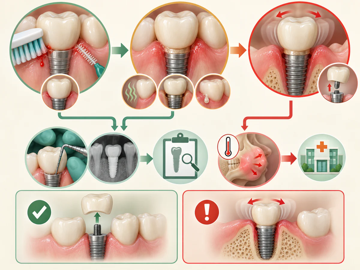
- Gum bleeding when brushing, when using an interdental brush, or on contact. This is the earliest and most useful warning sign.
- Redness, swelling or tenderness of the gum around the implant crown.
- Discharge of pus (suppuration), a sign of more marked active inflammation.
- Discomfort, pain or a persistent bad taste in the implant area. Pain is not always present, which makes the other signs all the more important.
- Gum recession exposing the neck of the implant, or a change in appearance.
- Implant mobility, that is, an implant that moves.
On this last point, we need to be clear. Mobility of the implant body reflects complete loss of osseointegration, and therefore failure of the implant (Berglundh et al. 2017; Schwarz et al. 2018). It is a late sign, not an early warning. An implant that moves does not “tighten back up” and does not re-integrate. The whole purpose of monitoring is precisely to intervene well before that stage, at the bleeding and inflammation phase, while the situation is still reversible or manageable.
Pain around an implant, bleeding that does not go away, or suppuration all warrant a prompt appointment. In the event of acute pain, significant swelling or fever, seek prompt care; our guide to what to do right away in a dental emergency explains the next steps.
What are the risk factors for peri-implantitis?
Not all patients are exposed in the same way. According to the review by Schwarz et al. (2018), the best-supported risk indicators are:
- a history of periodontitis;
- poor plaque control;
- the absence of regular maintenance.
The first point is often underestimated. Someone who has already lost bone around their natural teeth because of periodontitis remains more exposed around their implants. If you are unsure about the nature of your past gum problems, the article on the difference between periodontitis and gingivitis can help you situate your own profile.
Smoking and diabetes are also identified as risk factors by the EFP (Herrera et al., 2023). Some nuance is needed, however: in the review by Schwarz et al. (2018), the level of evidence for these two factors is judged inconclusive. They deserve to be taken into account in individual assessment, without being presented as certain, standalone causes.
In France, the HAS (2024) translates this logic into its care pathway: it explicitly recognizes mucositis and peri-implantitis, and sets as a prerequisite before implant placement a healthy mouth, free of active disease and with good oral hygiene. It is the same requirement we apply at the practice: treat first what needs treating, then place.
How can implant failure be prevented? The role of maintenance
Prevention rests less on the surgery itself than on what happens afterwards.
Peri-implant maintenance, the central measure
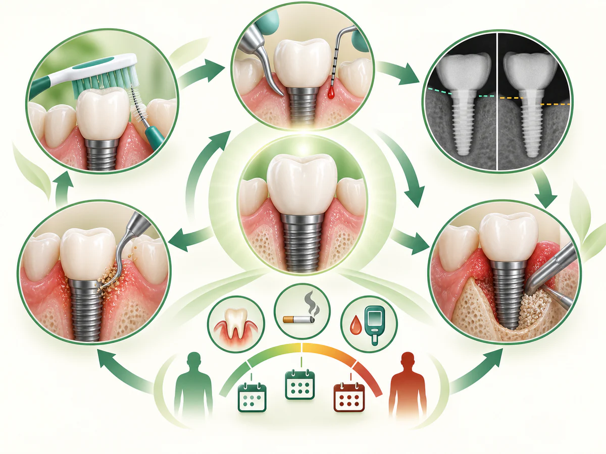
The EFP S3 guideline (Herrera et al., 2023) places maintenance, or supportive peri-implant care, at the heart of prevention: periodic professional removal of biofilm and reinforcement of the patient’s hygiene, with suitable tools such as interdental brushes. The HAS (2024) similarly describes a post-treatment follow-up phase combining professional maintenance, provided by the practitioner, and personal maintenance, carried out by the patient.
There is no universal recall interval that suits everyone. The follow-up rhythm, often a matter of a few months, is adapted to your level of risk. Someone with a history of periodontitis and a smoking habit will be followed more closely than someone at low risk. For everyday habits, the article on daily dental hygiene sets out the method and frequency to apply around implants as well as natural teeth.
The role of clinical equipment in monitoring
At the practice in Kénitra, follow-up of an implant relies on clinical examination, gentle probing and imaging. Radiography, most often a periapical X-ray and in some cases a Cone Beam scan (cone beam computed tomography), makes it possible to objectively assess the state of the peri-implant bone and to document any bone loss over time, an element that the definition of peri-implantitis makes indispensable. This imaging is also used beforehand, to plan placement and, where relevant, to assess a prior bone reconstruction when bone volume is insufficient. Monitoring only means something when compared with a baseline, hence the value of a continuous imaging record.
What to do if you suspect a problem with your implant
The course of action is simple and comes down to a few principles.
- Do not wait for pain to appear. Bleeding often comes before discomfort. Bleeding around an implant is already reason enough for an appointment.
- Do not self-treat with prolonged mouthwashes or antibiotics taken without advice. This can mask the signs without acting on the cause.
- Keep up your hygiene without irritating the area, and report any recent change.
- Come in for an assessment including examination, probing and, if necessary, a radiograph, in order to distinguish reversible mucositis from early peri-implantitis.
The earlier the care, the more conservative it is. Conversely, established peri-implantitis with bone loss calls for heavier interventions and its outcome is less predictable.
In summary
A dental implant can fail, and it often gives warning first. The clinical message fits in one sentence: watch for bleeding, treat early, do not wait for mobility. Peri-implant mucositis is reversible when the biofilm is controlled; peri-implantitis, which affects the bone, is not spontaneously reversible (World Workshop 2017 consensus). A history of periodontitis, plaque control and the regularity of maintenance are the most decisive levers (Schwarz et al. 2018; EFP 2023). Well-conducted implant treatment combines an indication set in a healthy mouth, as the HAS (2024) points out, and organized follow-up over time.
Frequently asked questions
My implant bleeds when I brush my teeth: is that serious?
Can an implant that moves be saved?
Is peri-implantitis common?
I have had periodontitis before: am I more at risk?
How often should I see the practitioner after an implant is placed?
Sources
Medical references consulted for this article.
- 1Berglundh et coll., rapport de consensus du World Workshop 2017 sur les maladies péri-implantaires, J Clin Periodontol 2018 (PubMed)
- 2Schwarz et coll., Peri-implantitis, World Workshop 2017, J Clin Periodontol 2018 (PubMed)
- 3Derks et Tomasi, revue systématique de l'épidémiologie péri-implantaire, J Clin Periodontol 2015 (PubMed)
- 4EFP, recommandation S3 sur la prévention et le traitement des maladies péri-implantaires (Herrera et coll., 2023)
- 5HAS (France), Implants et prothèses dentaires : avis et prise en charge des maladies péri-implantaires, 6 nov. 2024
- 6Chrcanovic et coll., survie des implants posés sur des sites d'implants perdus, Clin Oral Implants Res 2017 (PubMed)
- 7Shenoy et coll., échec implantaire et techniques de dépose : revue de la portée, Frontiers in Oral Health 2025
Contents
Share