Being told that you “don’t have enough bone” for an implant is a common source of worry, and the first thing to know is reassuring: in the great majority of cases, insufficient bone volume is not a definitive refusal. Several solutions exist. The missing bone can be rebuilt with a graft or a sinus lift, or, in certain situations, the graft can be avoided by using short or tilted implants. The UFSBD patient information sheet puts it clearly: when the bone has resorbed, the situation is more complicated, but an implant often remains feasible, sometimes after a graft intended to improve the receiving site. The right choice is made after a precise examination that measures the volume actually available. This article goes through each option, with its benefits, its limits and its contraindications.
Why does bone come to be lacking for an implant?
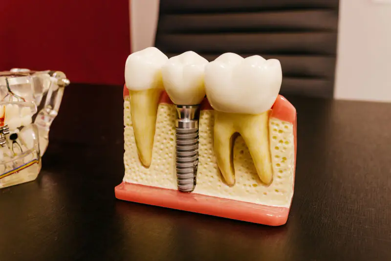
A dental implant is an artificial root anchored in the jawbone. To be stable and durable, it needs a sufficient volume of bone around it, in height as well as in thickness. When that volume is lacking, direct placement becomes difficult, or even impossible without prior preparation.
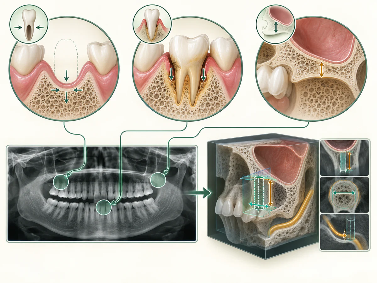
The most common cause is the bone resorption that follows the loss of a tooth. The alveolar bone, the bone that surrounded the root, is no longer stimulated by chewing and gradually recedes. A meta-analysis published in the Journal of Clinical Periodontology (Couso-Queiruga et al., 2021) quantified this phenomenon after spontaneous healing of a socket: the loss is measured in millimeters, above all in width, and occurs mainly during the first months following the extraction. Its extent varies from one person to another and according to the tooth concerned. The UFSBD sheet also lists this “bone loss at the site of the missing tooth” among the direct consequences of an absent tooth.
Other factors can reduce the available volume:
- an infection or periodontal disease (advanced gum disease) that has destroyed the supporting bone;
- an old extraction, the bone having had time to resorb;
- in the posterior upper jaw, the proximity of the sinus, whose floor can descend when the molars are missing;
- a trauma or a lesion that has taken away part of the bony ridge.
This is why early care after an extraction often limits later difficulties. You will find the main stages set out in detail in our article on how a dental implant is placed, step by step.
If a tooth is soon to be removed, what can be done at the time of extraction to keep bone volume is explained separately.
How is bone volume assessed before an implant?
The assessment never rests on a simple clinical impression. It begins with an examination of the mouth and a standard radiograph, most often a panoramic X-ray. When these two-dimensional images are not enough to judge precisely the volume and the position of the neighboring structures, a three-dimensional examination is prescribed.
The role of the Cone Beam
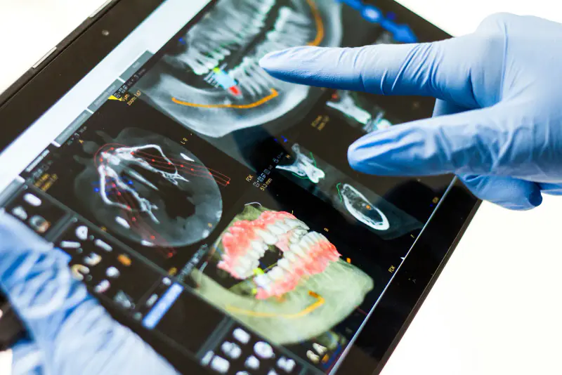
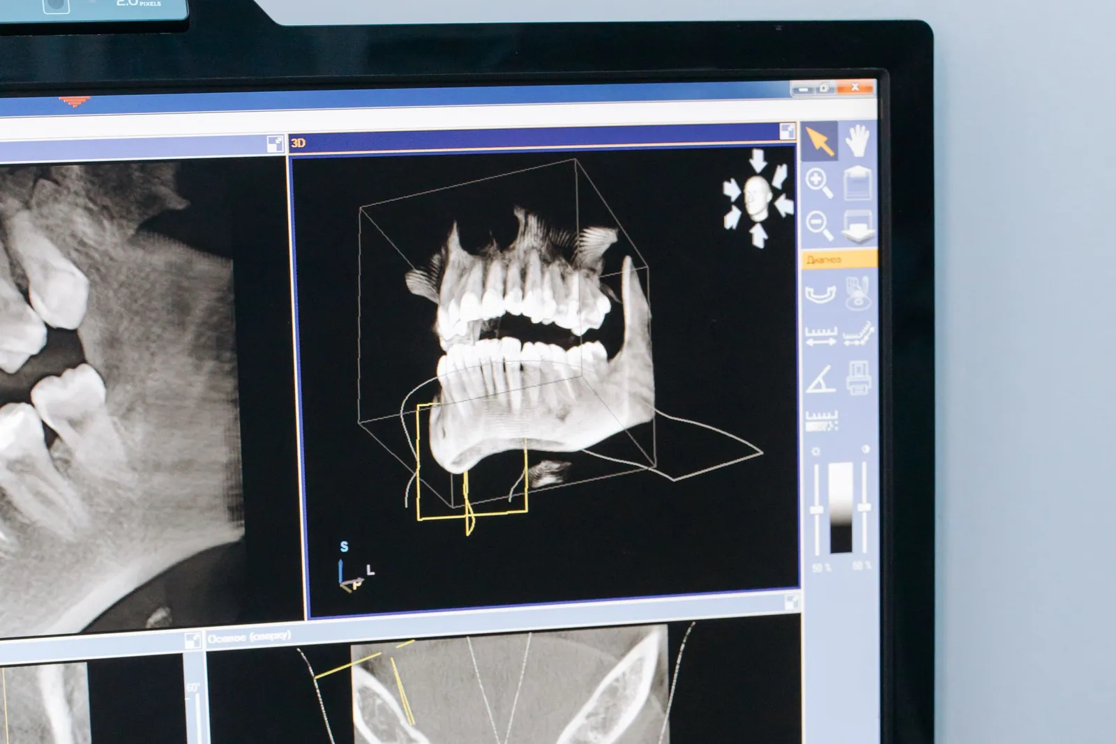
The Cone Beam, or cone beam computed tomography, provides a three-dimensional image of the bone. According to the memo on the appropriate use of the Cone Beam issued by the Assurance Maladie and the ADF (December 2021), which cites the Haute Autorité de Santé, this examination is indicated “for a pre-implant assessment and an estimate of the bone volume at the implant site”. The same source recalls that it is a second-line examination, justified when the clinical picture and two-dimensional radiology are not sufficiently informative and a three-dimensional image becomes indispensable.
In practice, the Cone Beam makes it possible to measure the available bone height and thickness to within a millimeter, to locate the inferior alveolar nerve in the mandible or the sinus floor in the maxilla, and to plan the procedure. At the practice we have a Durr Dental Cone Beam, which means this assessment can be carried out on site when it is needed. This examination is not routine: it is requested when the situation calls for it.
What is measured: height, width, distance to the nerve and the sinus
Four elements count, and they are read together.
- Bone height, between the top of the ridge and the obstacle above or below it: the sinus floor in the upper jaw, the nerve canal in the lower jaw. It limits the length of the implant. As an order of magnitude, a 2023 meta-analysis calls implants under 8 mm “short” and those of 8 mm or more “standard”, often 10 to 15 mm in the trials. Under the sinus, the Foundation for Oral Rehabilitation takes a remaining height of less than 4 to 6 mm as the usual indication for a sinus lift.
- Ridge width, from the cheek side to the tongue or palate side. The implant must be surrounded by bone on all sides, not just held in height.
- Distance to the nerve, in the lower jaw. The nerve that gives feeling to the lip and chin runs through the bone, and a margin is kept between it and the implant.
- The position of the future tooth. The implant is placed where the tooth needs to be, not just where bone remains.
So there is no single threshold. A shorter implant needs less height, a narrower implant less width, and every tooth plan is different. The course of the nerve also varies from person to person: a review of Cone Beam imaging (Weiss and Read-Fuller, 2019) reports that no minimum distance could be recommended for everyone at the front of the lower jaw, and that the anatomy is assessed case by case on the image.
What if the implant were placed without enough bone?
Placing an implant in too little bone exposes it to problems that a graft or the choice of another implant are precisely meant to avoid.
- Part of the implant is not covered by bone, and the gum can recede at that spot. A 2017 World Workshop review (Hämmerle and Tarnow) lists poor implant position and thin gum among the causes of bone and gum loss around implants.
- The implant may lack stability at the start, although this stability is needed for it to bond with the bone, a process called osseointegration. Bone density matters here as well as volume: denser bone holds the implant more firmly at placement.
- In the lower jaw, injury to the nerve while the bone is being prepared can cause altered feeling in the lip or chin (Greenstein and Tarnow, 2006).
- In the upper jaw, an implant longer than the available height would enter the sinus: this is what a sinus lift or a short implant avoids.
The medical factors examined alongside
Bone volume is not the only element studied. The UFSBD sheet specifies that good general health is a condition of success, because a chronic disease such as diabetes can slow healing and lead to failure. Smoking is assessed as well: a meta-analysis published in Medicina (2021) found in smokers a risk of implant failure roughly two and a half times higher than in non-smokers, along with more marked bone loss. These elements do not close the door on an implant, but they are discussed and taken into account in the treatment plan.
Rebuilding bone: graft, GBR and sinus lift
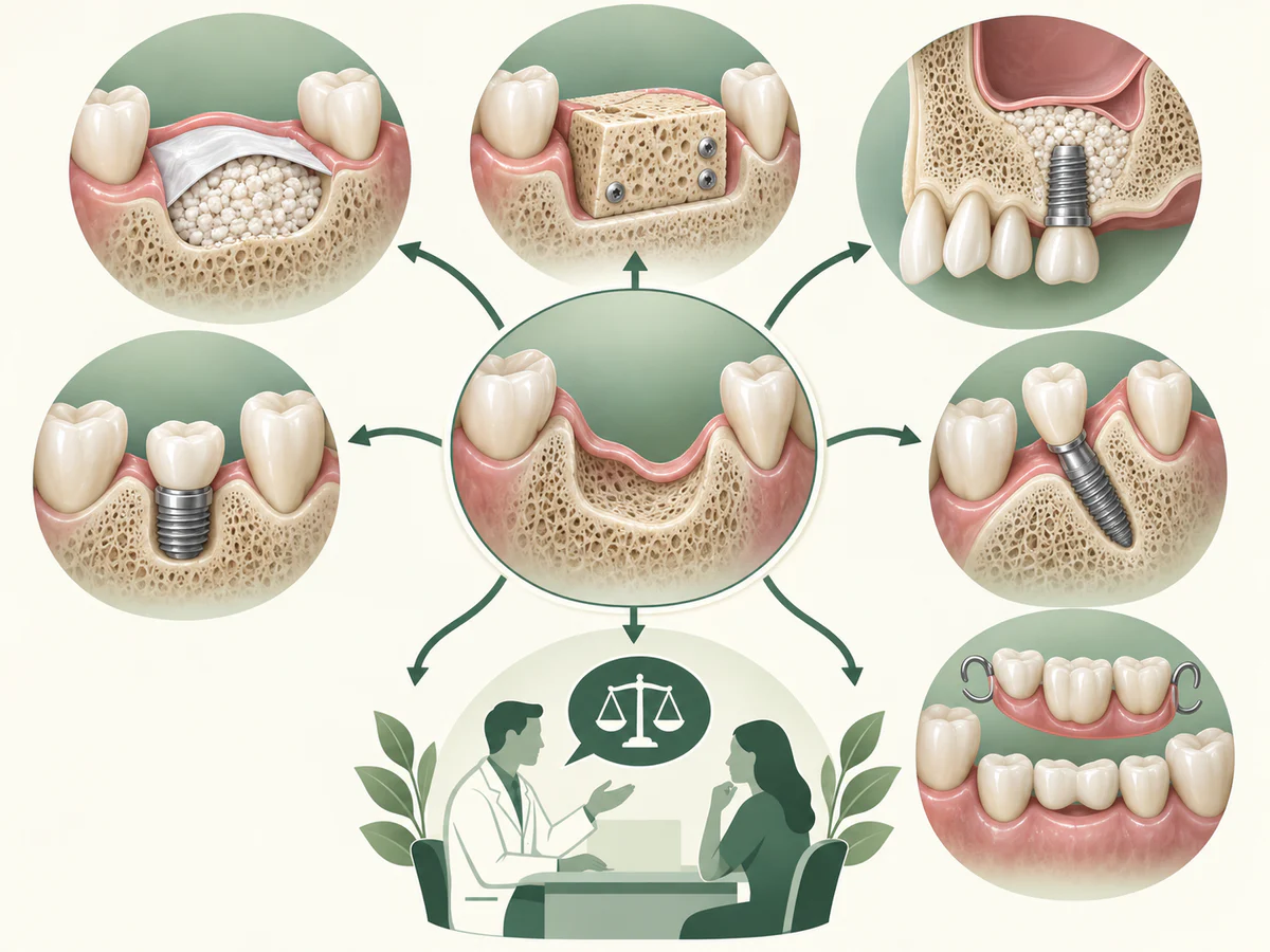
When the volume is insufficient, a first family of solutions consists in rebuilding the missing bone before or during placement of the implant. Rebuilding bone for dental implants is not the only route, however: a second family of solutions, described further on, avoids the graft in certain situations.
Bone grafting and guided bone regeneration
A bone graft consists in adding bone material in order to increase the volume of the ridge. Guided bone regeneration, or GBR, combines this material with a membrane that protects the site during healing. The 4th EAO consensus conference (2015) confirms that these augmentation techniques are valid options for making a site implantable. The UFSBD sheet indicates for its part that a graft may be carried out “to improve the site receiving the implant”.
Some nuance is needed, however. None of the recommendations consulted provides a universal success rate for grafting: the outcome depends on the type of graft, on the site and on patient-specific factors such as smoking or diabetes. The EAO 2015 also stresses that vertical augmentation, which aims to regain height, remains the most delicate and is accompanied by more complications. Announcing a fixed success percentage would therefore be misleading. The details of the procedure and of healing are described in our dedicated article on the bone graft before an implant: why, how and healing.
At the practice, piezosurgery and Choukroun’s PRF, a platelet concentrate prepared from the patient’s own blood, may be used depending on the indications to facilitate the bone work and the healing of the tissues. These are aids, not guarantees of results.
What happens during a bone grafting procedure
The aim of a bone grafting procedure is to give the implant enough bone all around it to remain stable over the long term. It is carried out under local anesthesia: the gum is opened over the ridge, the bone material is placed where volume is lacking, covered with a protective membrane in the case of GBR, and the gum is then closed over the site. Depending on the situation, the graft is done before the implant, with several months of healing before implant placement, or at the same time as the implant when enough bone remains to stabilize it. Moderate pain and swelling may follow, as after the placement of an implant. In other words, grafting is a procedure in its own right, with its own healing time, and when it is done before the implant it adds a stage to the overall treatment.
The sinus lift
In the posterior upper jaw, when the sinus has taken the space left by missing molars, its floor can be raised and the space filled with bone material. This is the sinus graft, also called a sinus lift. The EAO 2015 recognizes it as a valid option for addressing a lack of height in this area. The procedure then requires several months of healing before the implant is placed. How it unfolds in detail is set out in the article on the sinus lift: indications and procedure.
Avoiding a graft: short implants and angled implants
A graft is not always necessary. A second family of solutions consists in adapting the implant, or its orientation, to the existing bone volume.
Short implants
Short implants are designed to anchor in a reduced height of bone. This is the most important nuance to keep in mind: a graft is not always needed. The 4th EAO consensus conference (2015) reports that, in the posterior areas of the maxilla and the mandible, short implants lead to fewer complications than augmentation techniques, for comparable clinical results.
A meta-analysis with a follow-up of at least five years (2023), covering the atrophic posterior maxilla, found no statistically significant difference in survival between short implants and standard implants placed after a sinus lift. The short implants even showed less marginal bone loss and fewer biological complications. These data do not mean that the short implant is “better” in every circumstance. They indicate that it is a credible alternative, to be chosen case by case according to the available volume and the position of the tooth to be replaced.
Angled implants
In certain situations, tilting the implant makes it possible to bypass an area where bone is lacking, such as the sinus, and to make use of denser bone located elsewhere. This approach is recognized in implant practice. Three-dimensional imaging is decisive here in order to plan the axis of the implant safely.
Zygomatic implants, when almost no bone is left in the upper jaw
When the upper jaw is severely resorbed, long implants can be anchored higher up, in the cheekbone, called the zygomatic bone. A 2023 systematic review (Polido and colleagues) describes them as a graftless solution that can shorten treatment, for extreme bone loss, after other implants or a graft have failed, or after removal of part of the jaw. It also stresses their limits: significant surgical risks, specific complications such as sinusitis, and an indication to be assessed very strictly, reserved for experienced teams.
So this is not the usual alternative to a graft, but an option for specific cases. If it is being considered for you, the assessment sets out the conditions and who would carry it out. The more common choice, between a short implant and a sinus lift, is covered in detail in the article on sinus lifts.
Comparison table of the options when bone is lacking
| Option | Principle | When it is considered | Points to watch |
|---|---|---|---|
| Bone graft / GBR | Adding bone and a membrane to increase the volume | Ridge too thin or too low | Several months of healing; outcome varies with the site |
| Sinus lift | Raising the sinus floor and filling the space | Posterior maxilla, insufficient height | Prolonged healing before placement |
| Short implant | Implant suited to a limited height of bone | Posterior areas | Often fewer complications; chosen case by case |
| Angled implant | Modified orientation to bypass a deficit | Available bone offset from the ideal site | Three-dimensional planning required |
The choice between these options is discussed in consultation, after the assessment. None imposes itself from the outset: it is the objective measurement of the volume, cross-referenced with your medical situation, that guides the decision.
When an implant is not the chosen option
Sometimes, after the assessment, an implant is not the solution chosen, whether for medical reasons or by personal preference. Missing teeth can then be replaced by a bridge, supported by the neighboring teeth, or by a removable denture. Each solution has its own advantages and drawbacks, which are compared in our article on choosing between an implant, a bridge or a denture.
How long does it take, and what follows the procedure?
The time factor is essential to understand. The solidity of an implant rests on osseointegration, that is, the healing of the bone around the implant. The UFSBD sheet puts the average time at around a few months. When a graft or a sinus lift precedes placement, several additional months generally have to be allowed for. Some protocols allow a fixed temporary prosthesis fairly quickly, but the final prosthesis comes once healing has been achieved.
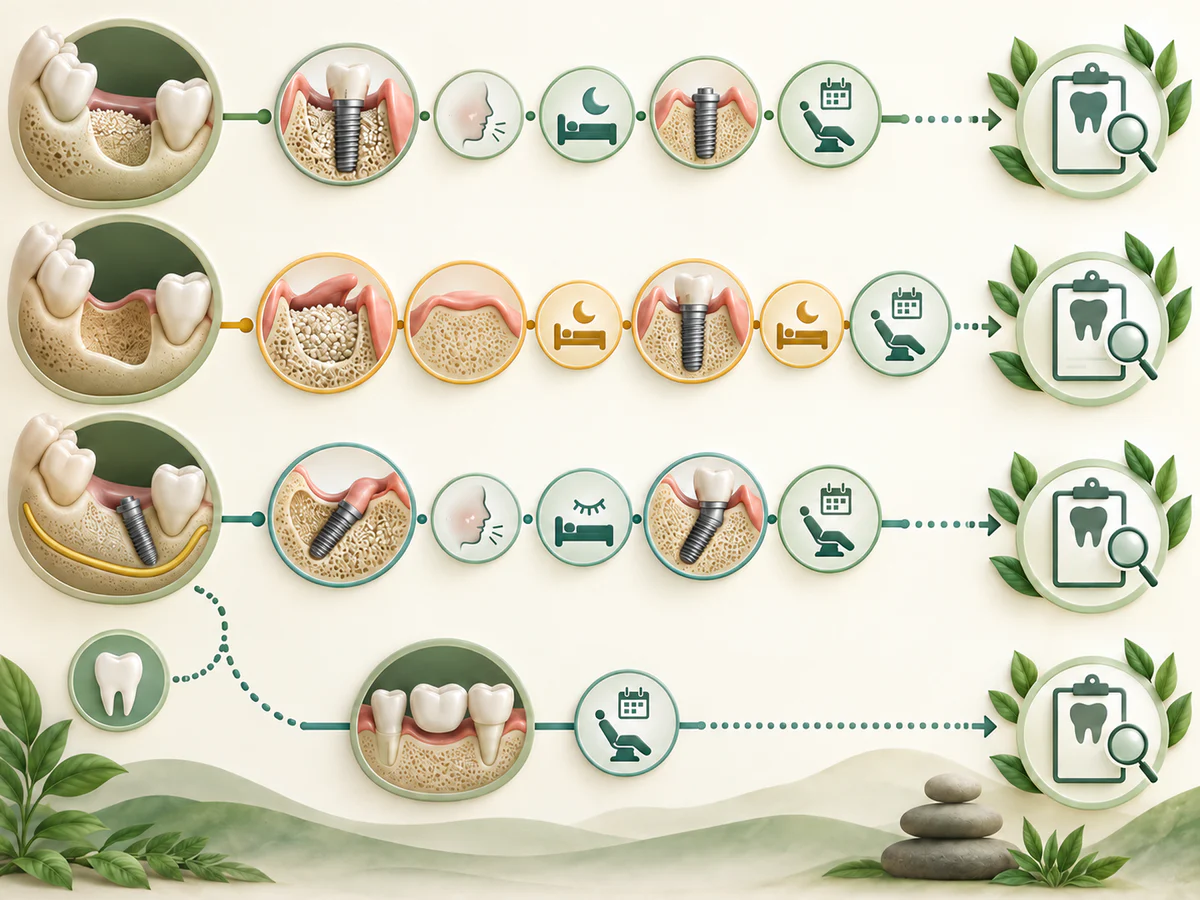
As for what follows, it is important to remain accurate. The procedure is carried out under local anesthesia. The UFSBD sheet indicates that moderate pain and inflammation may occur after the procedure; they are managed with the prescribed painkillers. Speaking of a “painless” or “risk-free” surgery would be inaccurate.
Finally, an implant is not permanently sheltered from problems. The consensus of the 2017 World Workshop (EFP/AAP) defines peri-implantitis as an inflammatory process of microbial origin leading to bone loss around the implant. We do not speak of “rejection” as with an organ transplant: a failure stems above all from a defect in osseointegration or from an infection, favored by smoking, poorly controlled diabetes or insufficient hygiene. An implant helps preserve the bone in the area, but marginal bone loss remains possible. To recognize the warning signs, see our article on implant failure and peri-implantitis: signs to watch for.
The reconstruction techniques offered at the practice are described on the Bone graft page; implant placement, once the bone has been evaluated, on the Dental implant page.
Key points to remember
As the UFSBD sheet recalls, nothing replaces a natural tooth, but the implant is the closest thing to it. A lack of bone is, most often, not an insurmountable obstacle. Several routes exist: rebuilding the bone with a graft, GBR or a sinus lift, or adapting to the existing volume with short or angled implants. Each option has its indications, its limits and its healing time. The decision rests on a clinical and radiological assessment, completed if necessary by a Cone Beam, and on taking your medical factors into account. It is this individual evaluation, and not a single rule, that makes it possible to propose the most appropriate solution.
To start with the words you may have been told, read What the assessment measures when you were told ‘there is not enough bone’.
Understanding the bone grafting materials
A bone graft for dental implants is a separate treatment stage when the existing bone cannot safely support the implant. The choice of bone grafting materials, the need for a membrane and the healing interval depend on the defect and the proposed restoration. Ask which material is planned and why, rather than assuming every graft follows the same method. Our guide to dental bone grafting before an implant explains the types of bone grafts and their limits in more detail.
Frequently asked questions
I've been told I don't have enough bone for an implant: is that a definitive refusal?
Is a bone graft always needed when bone is insufficient?
Is the procedure painful?
Does smoking prevent an implant from being placed when bone is limited?
How long do you have to wait before the crown is placed?
Does the jaw bone keep shrinking after tooth loss?
Sources
Medical references consulted for this article.
- 1UFSBD, Fiche patient « Un implant pour remplacer une dent », 2016
- 2Assurance Maladie (ameli.fr), Renforcer le bon usage du cone beam en odontostomatologie, 2022
- 3Assurance Maladie / ADF, Mémo « Le bon usage du cone beam », décembre 2021 (citant la HAS)
- 44e conférence de consensus de l'EAO, Clinical Oral Implants Research, 2015
- 5Couso-Queiruga et al., Post-extraction dimensional changes, J Clin Periodontol, 2021
- 6Mustapha, Salame, Chrcanovic, Smoking and Dental Implants, Medicina, 2021
- 7Berglundh et al., Péri-implantites, 2017 World Workshop (EFP/AAP)
- 8Implants courts versus comblement de sinus, méta-analyse à 5 ans, 2023
- 9Foundation for Oral Rehabilitation (FOR.org), Sinus lifting treatment guideline
- 10Weiss et Read-Fuller, Cone Beam Computed Tomography in Oral and Maxillofacial Surgery: An Evidence-Based Review, Dentistry Journal (2019)
- 11Hämmerle et Tarnow, The etiology of hard- and soft-tissue deficiencies at dental implants, J Clin Periodontol (2018), PMID 29926502
- 12Greenstein et Tarnow, The mental foramen and nerve: factors related to dental implant placement, J Periodontol (2006), PMID 17209776
- 13Polido et coll., Indications for zygomatic implants: a systematic review, International Journal of Implant Dentistry (2023)
Contents
Share