You were sitting in the dental chair. The dentist looked at an X-ray and said: ‘There is not enough bone.’ Perhaps the next steps were explained. Perhaps not. You went home with that statement, and it gradually came to mean ‘never’.
Let us take it word by word.
‘Not enough’: not enough for what?
Bone is not measured in isolation. It is measured in relation to a specific area and to the tooth being considered for replacement. Its height matters, but so do its thickness and its proximity to a nerve or the sinus. Until these elements are connected to a plan for the tooth, the statement remains incomplete.
‘Bone’: which bone, seen how?
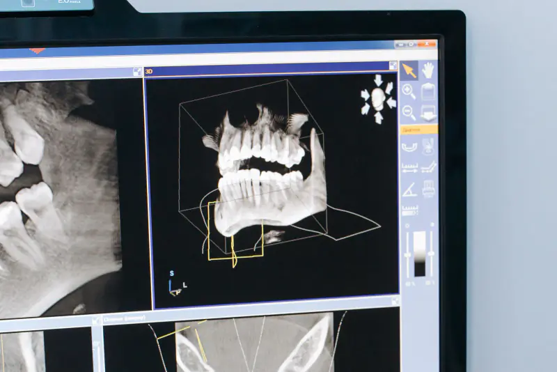
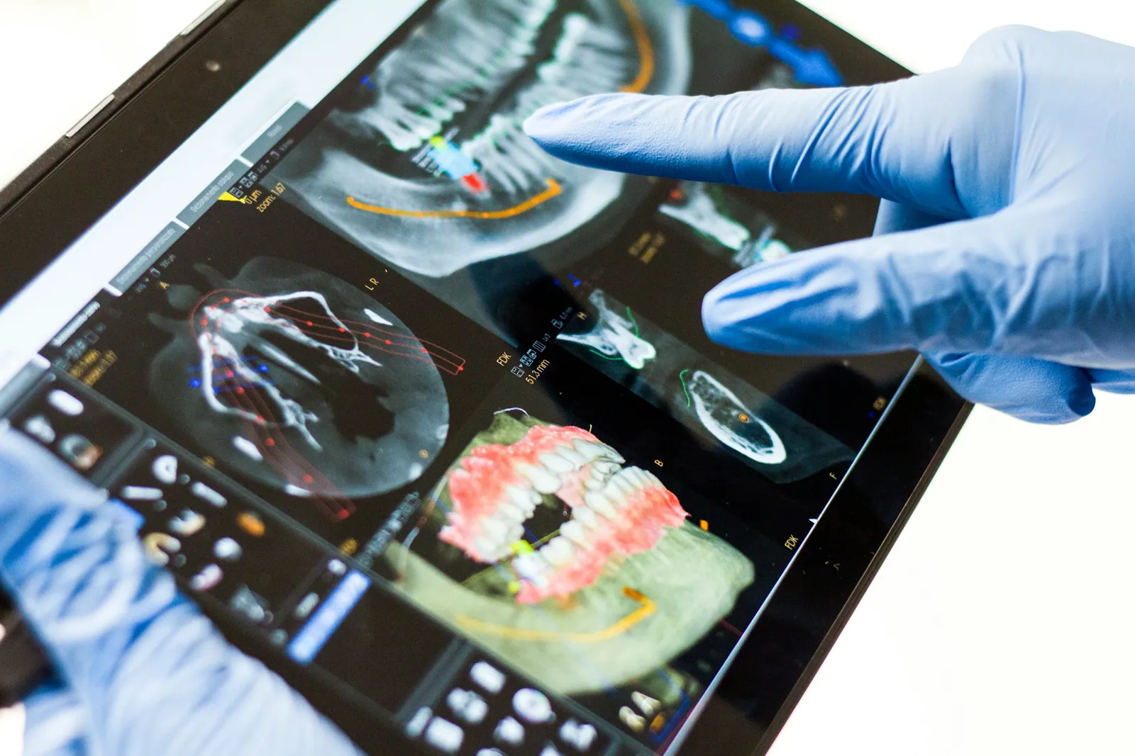
A panoramic X-ray provides a useful overview. It does not always show every dimension needed for a decision. When the volume needs to be defined in three dimensions, cone beam CT may be indicated. This examination is not automatic: it must answer a specific clinical question.
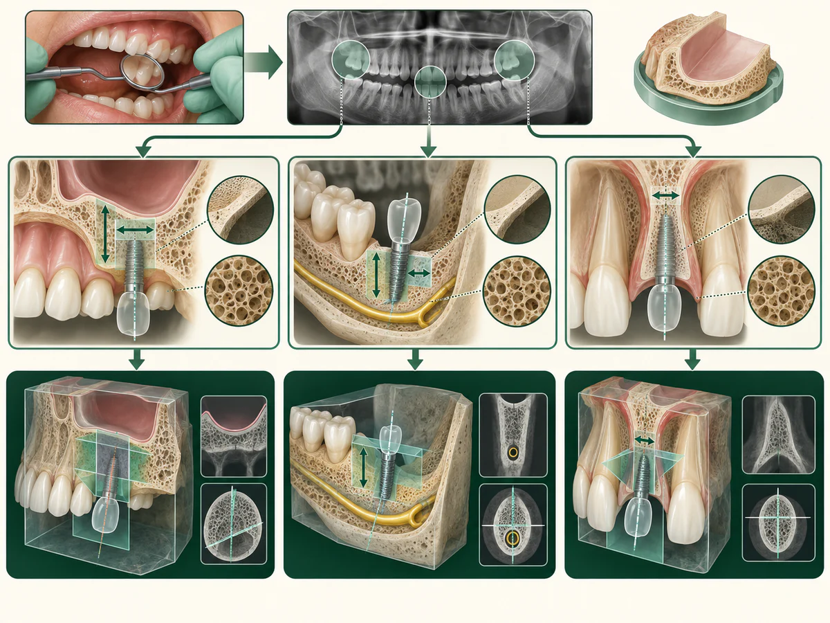
The assessment may confirm that the available volume is limited. That still does not decide the treatment. Instead, it points to the paths that may be discussed.
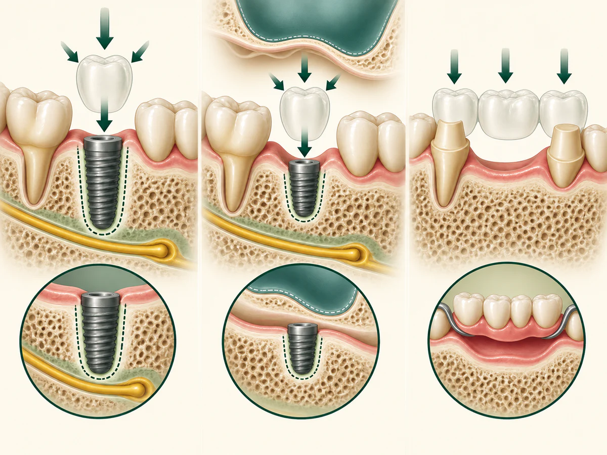
The first path is sometimes to rebuild the bone and then allow the area to heal before reassessing what comes next. At the back of the upper jaw, sinus floor augmentation may be among the techniques considered in some situations.
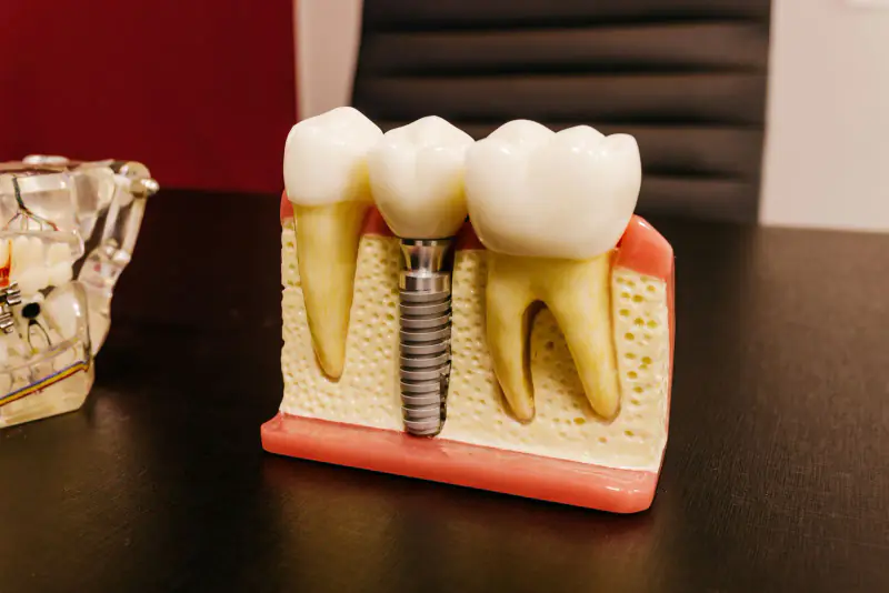
Another path does not involve an implant: a bridge supported by the neighbouring teeth, or a removable denture. These are not secondary solutions by definition. Their suitability depends on your mouth, your priorities and their maintenance. The examination may also lead to doing nothing for the time being.
A graft is therefore neither automatic nor a sign that someone was mistaken. It is an anatomical option that can only be discussed after the site has been measured. The article Dental implant without enough bone presents the techniques in detail; this page stays focused on your first question: what does the statement you heard mean?
How to check what you were told
You may leave with a doubt: is the bone loss really that significant? To clear it up, there is no need to suspect anyone. The answer is on your own image, because a measurement can be shown. You can ask to see, on your X-ray or your Cone Beam scan, where the implant would go and the bone that would surround it.
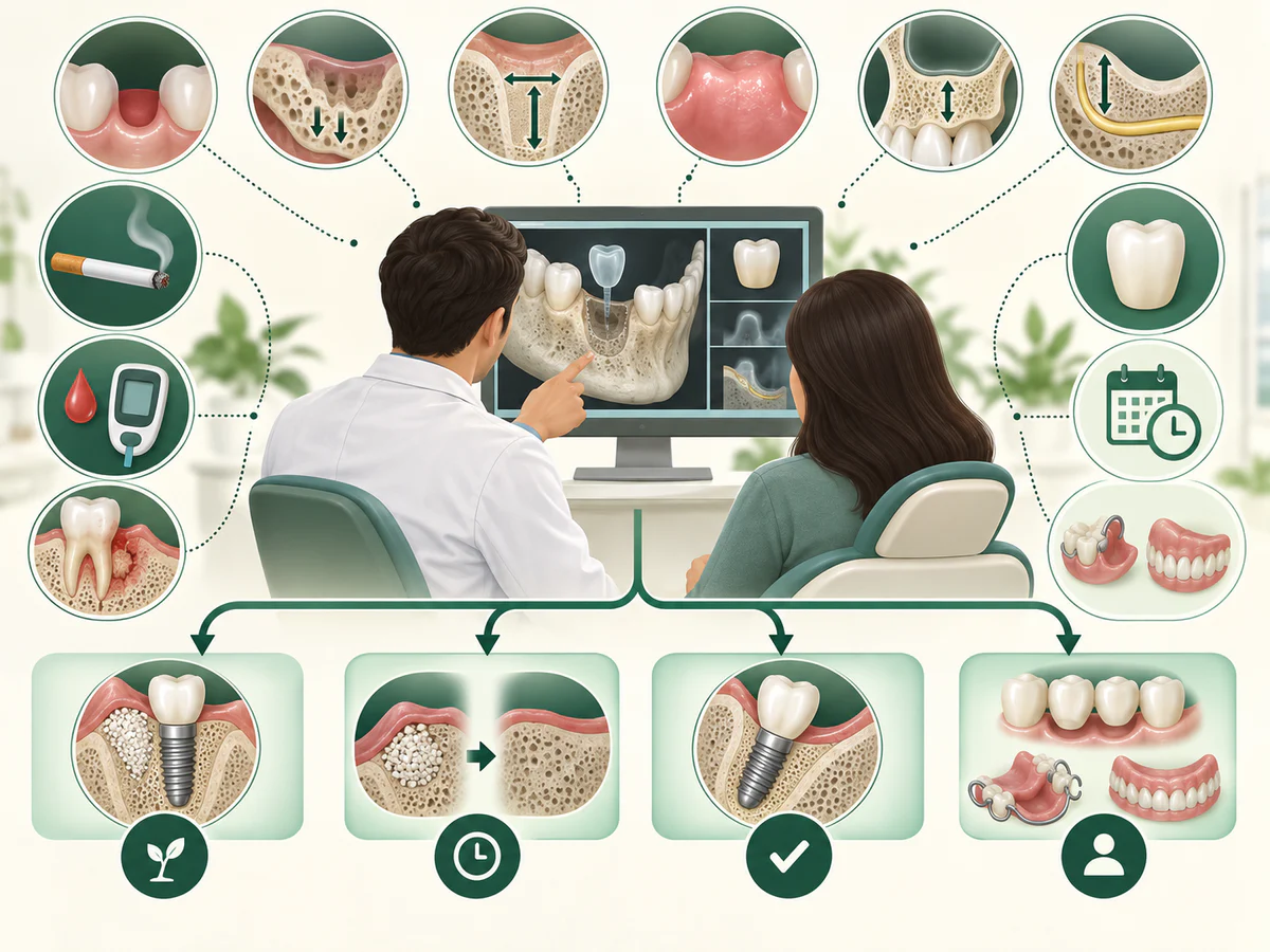
Three questions make the sentence checkable:
- “How much bone is there at this spot, in height and width, and how much would the planned implant need?”
- “Where will the final tooth be, and could the implant be placed differently, or be shorter?”
- “Without a graft, which options remain possible, and why are they not suitable?”
An answer with figures, shown on the image, lets you understand the proposal and compare it if you ask for another opinion. Why two practitioners can reach different conclusions is explained in graft or no graft: why two opinions can differ.
If the tooth has not yet been removed, what can be done at the time of extraction can also be discussed.
What the assessment should clarify
- What is happening: the height and thickness of the bone in the area concerned, along with the anatomical structures that must be respected.
- What needs prompt attention: rarely the lack of bone itself; pain, infection or swelling changes the priority.
- The reasonable options: bone reconstruction, a solution without an implant, or monitoring, depending on the examination.
- The steps: their order and the healing times when reconstruction is being considered.
- The plan: if treatment is being considered, the information needed to describe the steps and establish the cost after the evaluation.
The assessment is carried out by Dr Fatima Azelmat, a dental surgeon in Kenitra. It connects the bone measurements with the gum, bite and planned final tooth. The Bone grafting in Kenitra page describes the main techniques; none can be chosen from a statement alone. Scheduling an assessment does not commit you to any treatment.
If the measurement allows it, the next step is called implant placement, step by step; if not, other routes exist. The assessment measures before naming anything.
Frequently asked questions
Does ‘not enough bone’ mean an implant is impossible?
Is a panoramic X-ray enough to measure the bone?
Is a graft always necessary when bone is lacking?
Can I get a price before the bone is measured?
Sources
Medical references consulted for this article.
Contents
Share