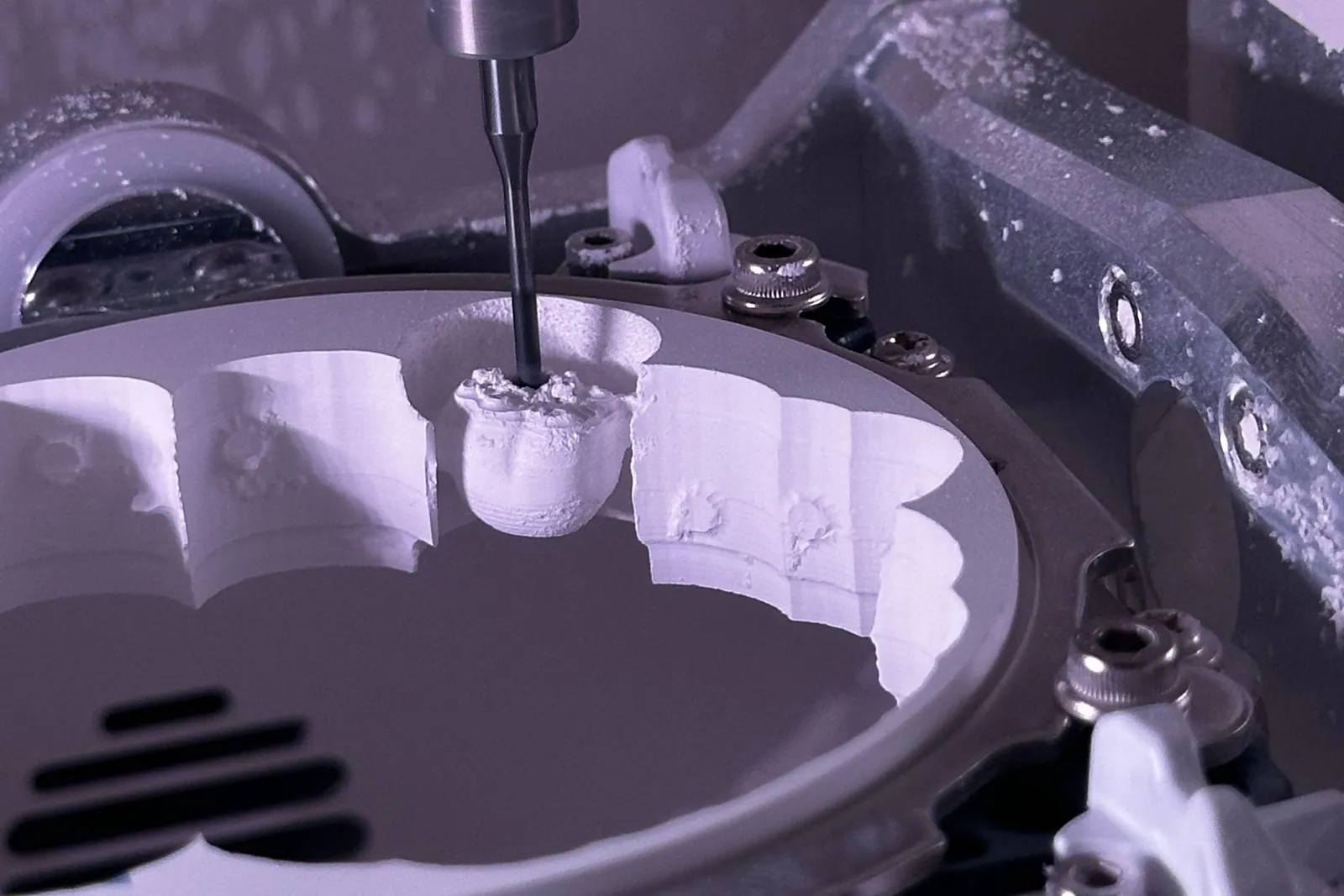
Computer-aided design and computer-aided manufacturing is called CAD/CAM for short. In French, it is known by the acronym CFAO. It is a set of tools used to design a dental restoration on a computer. The restoration is then produced by milling or printing.
In practice, it replaces some of the manual steps of impression-taking and fabrication with a digital workflow. It is a technology, not a treatment in itself. It is used to produce crowns, veneers, inlays/onlays (partial restorations that fill or cover part of a tooth), and bridges. But the clinical indication and the practitioner’s work determine the result, not the machine.
As a dental surgeon in Kenitra, I offer here a factual, measured description of CAD/CAM. It covers:
- what CAD/CAM is;
- how the workflow runs, from the scan to the restoration;
- which materials are used;
- what published studies say about the fit and survival of digital restorations;
- where its limits lie.
Our practice uses a digital CAD/CAM workflow. Every statement is backed by a source you can check. No figure is presented as an individual guarantee. These are study data, to be read as probabilities.
What is CAD/CAM in dentistry?
CAD/CAM brings two families of tools to dental prosthetics, the field of crowns, bridges and other replacement pieces. The first is computer-aided design, or CAD: designing the restoration on a screen. The second is computer-aided manufacturing, or CAM: the automated production of the piece.
CAD/CAM does not create the tooth. It helps design and mill a restoration. The practitioner must still fit it, adjust it, and bond or cement it.
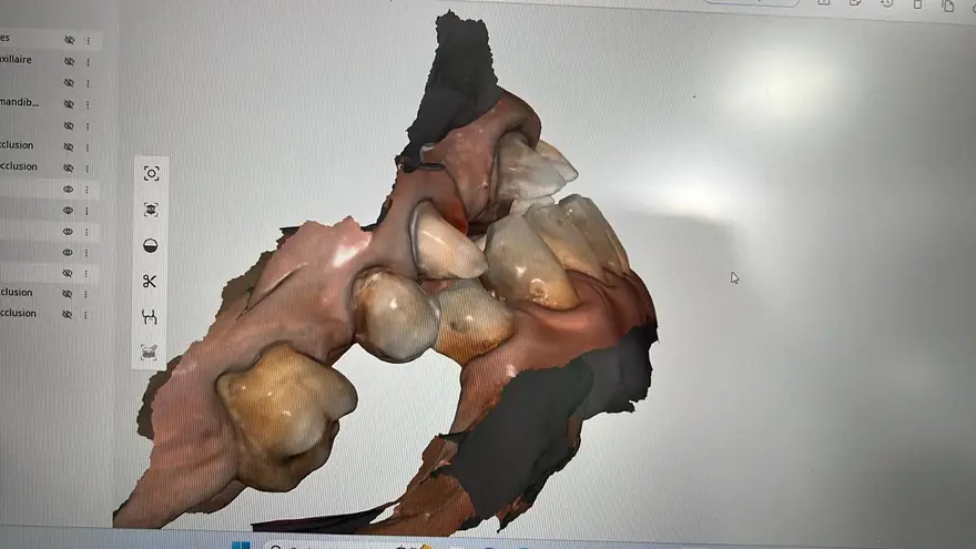
A review published in Bioinformation (Maiti and colleagues, 2024) describes the process in three stages:
- “the first step is to capture the dental anatomy with an optical scan to create a digital impression;”
- “the second uses CAD software, where the practitioner designs the restoration on a computer;”
- “the last implements CAM, where the design is sent to a milling machine or a 3D printer.”
These three links (scanning, design, manufacturing) form the digital chain.
There are two main ways to manufacture the piece:
- Subtractive milling starts from a block of material, such as ceramic. A milling unit carves the restoration out of this block.
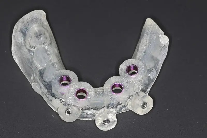
- 3D printing is an additive process. It builds the piece layer by layer.
The two exist side by side and do not serve the same purposes. Milling remains the reference for ceramic crowns and veneers. Printing is developing mainly for models, guides, and provisional (temporary) restorations.
The digital workflow: from scan to restoration
The CAD/CAM workflow replaces the putty impression with a digital scan. It then moves on to design and manufacturing. In outline, it involves the following steps:
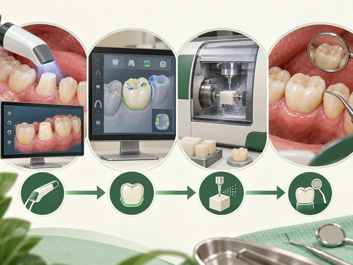
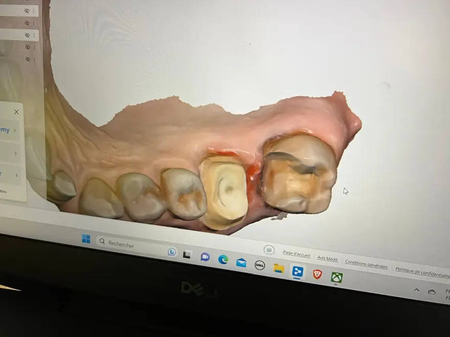
- The digital impression. An intraoral scanner is a handheld optical camera used inside the mouth. It records the shape of the prepared tooth, the neighboring teeth, and the occlusion (the way upper and lower teeth meet). It then produces a 3D model. Our article on digital impressions with an intraoral scanner details this step, its indications, and its limits.
- Design (CAD). On this model, the practitioner or the dental technician designs the restoration. This covers its shape, contact points, occlusion, and margin lines (where the restoration meets the tooth). The software proposes a shape, which is then adjusted to the case.
- Manufacturing (CAM). The design is sent to a milling machine or a printer. The piece is milled from a block, then finished. Finishing means staining, glazing, and, for certain ceramics, crystallization firing in a furnace.
- Try-in and placement. The restoration is tried in the mouth. Its fit and occlusion are checked and adjusted. It is then bonded or cemented.
This workflow can be fully digital, with a direct chairside scan (made at the dental chair). It can also be hybrid: a conventional impression, then a scan of the model at the laboratory. The distinction matters for fit, as we will see below.
Materials used in CAD/CAM restorations
CAD/CAM does not define a material. It defines a way of manufacturing. Several families of blocks are available. Each has its own trade-off between esthetics (appearance) and strength.
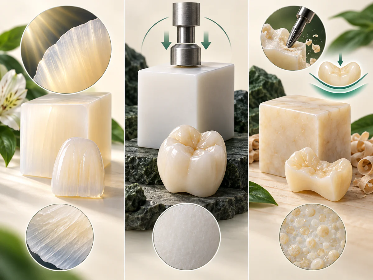
- Lithium disilicate is a reinforced glass-ceramic. It is esthetic and strong enough for many single crowns on front teeth and premolars. The review by Maiti and colleagues (Bioinformation, 2024) credits it with a flexural strength above 350 MPa. Flexural strength measures how much bending a material can resist before it breaks.
- Zirconia (zirconium oxide) is much stronger. The same source cites a flexural strength of 500 to 1200 MPa. This makes it suitable for areas under heavy chewing load. It also suits patients who clench or grind their teeth.
- Hybrid ceramics and millable composites are easy to mill and show intermediate behavior.
The material is chosen exactly as for a conventional restoration. The choice depends on:
- the tooth’s position;
- chewing forces;
- esthetic expectations.
Our articles on ceramic and zirconia crowns and ceramic dental veneers cover this topic in more depth. CAD/CAM is the shaping tool. The material and the indication remain the true clinical variables.
What the literature says about fit
The argument most often made for digital workflows is the precision of the fit. Fit means the marginal and internal adaptation of the restoration. This is how closely it matches the tooth at its edges and on the inside. The data broadly point in that direction, but with nuances.
A systematic review gathers and weighs all the studies published on one question. One such review came from Arcuri and colleagues (Journal of Biological Regulators and Homeostatic Agents, 2019). It covered all-ceramic restorations evaluated in vivo, meaning in patients’ mouths. Its conclusion: “CAD/CAM restorations obtained with an intraoral scanner show better marginal and internal fit than those obtained from a conventional impression digitized at the laboratory.” In other words, for fit, the direct digital workflow is a valid alternative to the indirect workflow, and even a favorable one.
A more recent umbrella review, which pools several reviews, came from Singh and colleagues (Cureus, 2025). It confirms that intraoral scanners offer clinically acceptable accuracy for single-unit and short-span restorations. However, it adds a clear caveat. Their accuracy decreases in complex cases, particularly full arches and edentulous situations (jaws without teeth). In these cases, “conventional or hybrid workflows may remain preferable.” So the superiority of digital is not universal. It depends on the span (how many teeth the restoration covers) and the clinical context.
What the literature says about survival
A good fit does not guarantee longevity. On this point, there is substantial follow-up data on CAD/CAM restorations, especially single-unit pieces.
For chairside restorations, the review by Fasbinder (Journal of the American Dental Association, 2006) looked at restorations produced with the CEREC system. It reported a survival probability “of approximately 97% at five years and 90% at ten years.” The main mode of failure was ceramic fracture.
Over the very long term, a case series by Otto (International Journal of Computerized Dentistry, 2017) followed CEREC 1 inlays and onlays. It observed survival of approximately 87.5% at up to 27 years. This figure is based on Kaplan-Meier analysis, a standard statistical method for estimating survival over time. The author describes the result as “highly acceptable” for restorations placed in private practice.
For lithium disilicate specifically, the review by Pieger, Salman, and Bidra (Journal of Prosthetic Dentistry, 2014) looked at single crowns. It reported cumulative survival of approximately 100% at two years and 97.8% at five years.
For all-ceramic CAD/CAM bridges, the source is a systematic review and meta-analysis by Saravi and colleagues (Materials, 2021). A meta-analysis combines the results of several studies with statistics. It reported five-year survival in the range of 89.7% to 91.1%. The leading cause of failure was secondary caries, meaning new decay at the edge of the restoration. The most frequent complication was chipping of the veneering ceramic (the outer ceramic layer).
| CAD/CAM restoration type | Reported survival | Source |
|---|---|---|
| Chairside restorations (CEREC) | ≈ 97% at 5 years, ≈ 90% at 10 years | Fasbinder, JADA 2006 |
| CEREC 1 inlays/onlays | ≈ 87.5% at up to 27 years | Otto, Int J Comput Dent 2017 |
| Lithium disilicate crowns | ≈ 97.8% at 5 years | Pieger et al., J Prosthet Dent 2014 |
| All-ceramic bridges | ≈ 89.7 to 91.1% at 5 years | Saravi et al., Materials 2021 |
These figures are survival rates drawn from studies. They are not a promise for any given restoration. Longevity depends on the manufacturing technology, but just as much on:
- the supporting tooth;
- the occlusion;
- oral hygiene;
- the patient’s habits.
The single-visit restoration: possible, but not routine
One advantage highlighted for chairside CAD/CAM is the single visit. Certain restorations can be designed and milled within the same visit. This means no putty impression and no temporary restoration while waiting for the laboratory. This is real, and the survival data above partly concern this workflow.
But it is neither automatic nor suited to every case. Whether a single visit is feasible depends on:
- the type of restoration: a single back-tooth crown is not a complex bridge;
- the chosen material: some ceramics require firing or staining that lengthens the timeline;
- esthetic demands: the front teeth often call for finer prosthetic work.
Presenting the single-visit restoration as a standard that applies everywhere would be an overpromise. It is a useful option in selected cases, not a general rule.
The limits of CAD/CAM
Honest information means naming this technology’s limits.
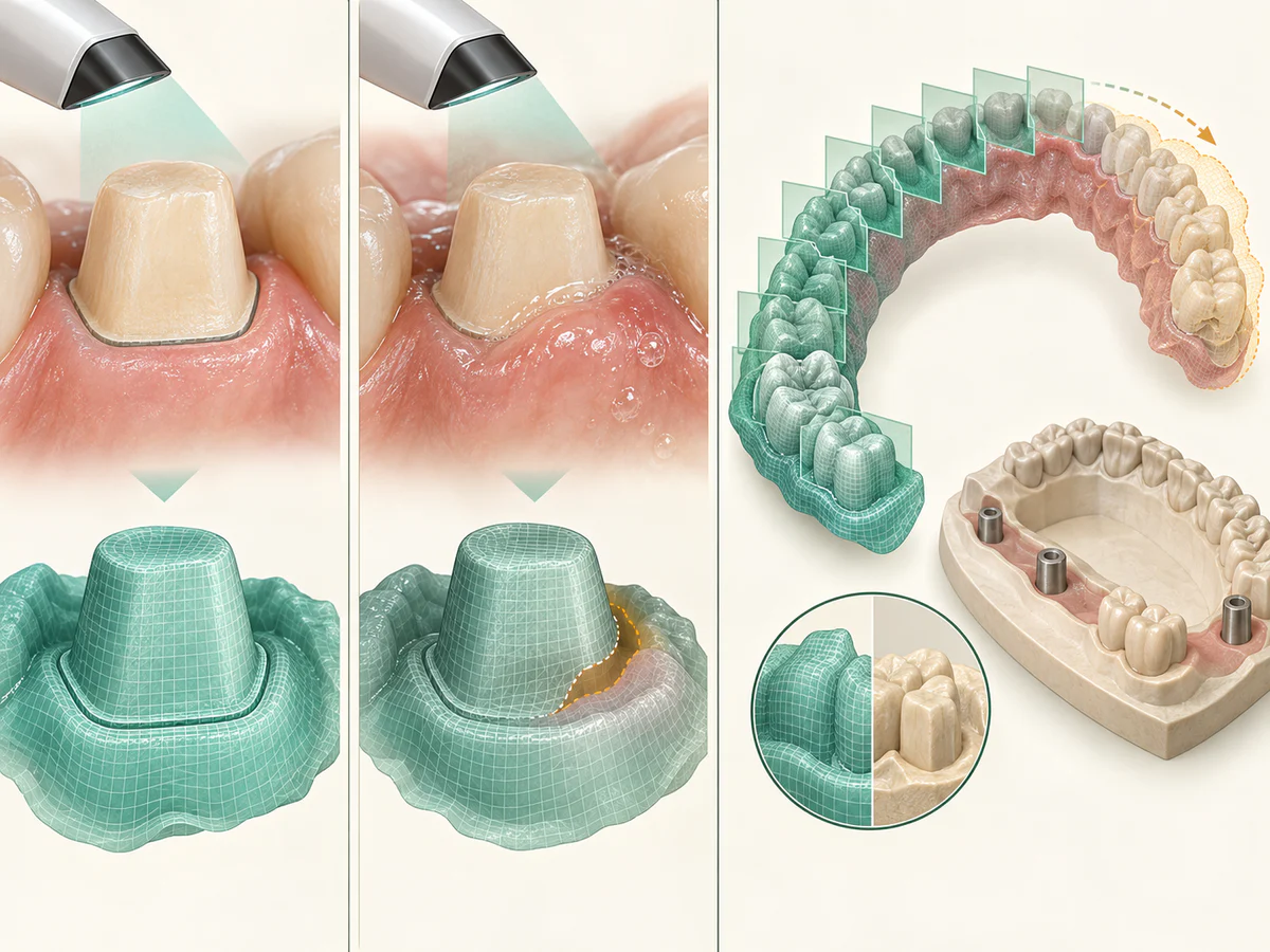
First, digital workflows do not remove clinical constraints. The same problems degrade a scan just as they degrade a conventional impression:
- a preparation margin (the edge of the shaped tooth) that is hard to see;
- bleeding gums;
- excessive moisture.
The quality of the result depends above all on the preparation and on control of the working environment.
Second, superiority is not systematic. The umbrella review by Singh and colleagues (Cureus, 2025) highlights the limits of scanners on full arches and edentulous cases.
The systematic review and meta-analysis by Aswal and colleagues (Cureus, 2023) compared CAD/CAM crowns and bridges with conventional restorations. The CAD/CAM pieces were made of lithium disilicate and zirconia. The review concluded that biological, technical, and esthetic outcomes were “broadly similar.” Yet in their analysis, the authors observed “more biological, technical, and esthetic complications in the CAD/CAM group than in the conventional group.”
The authors consider that CAD/CAM techniques “still need to evolve” to surpass conventional techniques. This nuance matters. CAD/CAM is a capable, convenient tool. It is not a guarantee of clinical superiority over well-executed conventional work.
Finally, even a restoration designed digitally to perfection still ages and wears. It remains subject to the same complications as any restoration:
- caries (decay) at the margin;
- fracture;
- debonding (the piece coming unstuck).
Regular follow-up and good oral hygiene remain essential.
In summary
CAD/CAM is a digital chain of scanning, design, and manufacturing. It makes it possible to design and mill ceramic dental restorations, from lithium disilicate to zirconia. The literature indicates:
- marginal fit that is at least comparable, and often better, with the direct digital workflow for single-unit restorations (Arcuri et al., 2019);
- favorable medium- and long-term survival (around 97% at 5 years for chairside restorations according to Fasbinder, 2006);
- in selected cases, the possibility of a single-visit restoration.
But this is not universal superiority:
- scanners have their limits on full arches;
- complications do occur (Aswal et al., 2023);
- no lifespan is ever guaranteed.
CAD/CAM is a tool in the service of the indication and of follow-up. It is not a substitute for either.
Frequently asked questions
What is CAD/CAM in dentistry?
Does a CAD/CAM restoration fit better than a conventional one?
How long does a CAD/CAM crown last?
Can you really get your restoration in a single visit?
Which materials are used in digital prosthetics?
Is CAD/CAM always superior to conventional work?
Sources
Medical references consulted for this article.
- 1Maiti N et al., Application of CAD-CAM in Dentistry, Bioinformation, 2024 (définition, flux en trois temps, matériaux)
- 2Arcuri L et al., Fit evaluation of CAD/CAM fabricated all-ceramic restorations based on direct and indirect digitalization in vivo (revue systématique), J Biol Regul Homeost Agents, 2019
- 3Singh S et al., Accuracy and Clinical Performance of Intraoral Scanners Compared to Conventional and Extraoral Impressions (revue parapluie), Cureus, 2025
- 4Fasbinder DJ, Clinical performance of chairside CAD/CAM restorations, Journal of the American Dental Association, 2006
- 5Otto T, Up to 27-years clinical long-term results of chairside Cerec 1 CAD/CAM inlays and onlays, International Journal of Computerized Dentistry, 2017
- 6Pieger S, Salman A, Bidra AS, Clinical outcomes of lithium disilicate single crowns and partial fixed dental prostheses (revue systématique), Journal of Prosthetic Dentistry, 2014
- 7Saravi B et al., Clinical Performance of CAD/CAM All-Ceramic Tooth-Supported Fixed Dental Prostheses (revue systématique et méta-analyse), Materials, 2021
- 8Aswal GS et al., Clinical Outcomes of CAD/CAM (Lithium disilicate and Zirconia) Based and Conventional Full Crowns and Fixed Partial Dentures (revue systématique et méta-analyse), Cureus, 2023
Contents
Share