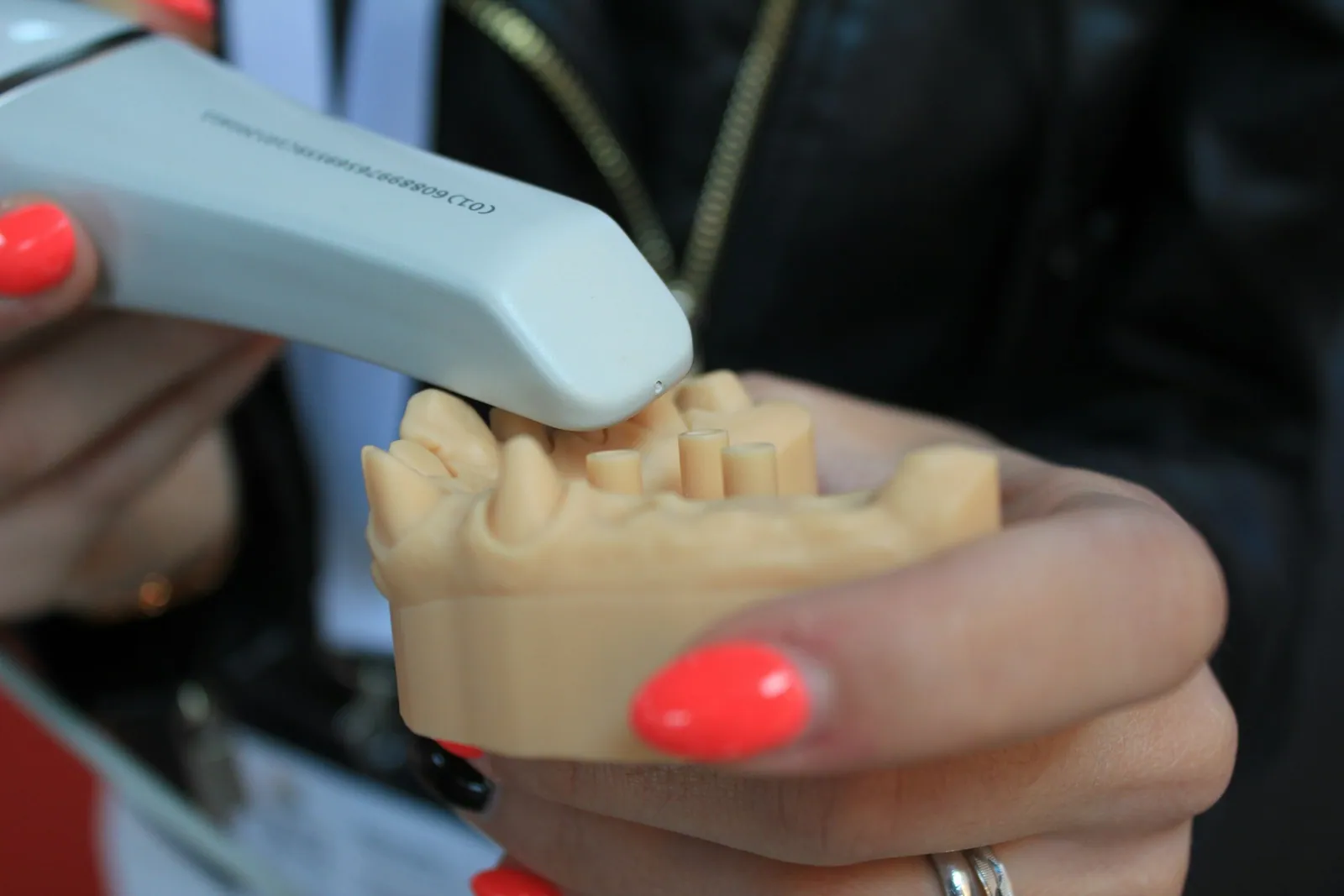
A digital impression records the shape of the teeth and gums no longer with a paste placed in an impression tray, but with a small optical camera called an intraoral scanner. Passed along the dental arches, it captures thousands of images that are assembled into a three-dimensional model displayed on screen in real time. The result is a digital file, the optical impression, which replaces the plaster cast poured from a conventional impression. At the practice in Kénitra, this is the method we use for many restorations, and this article describes how it works, what it offers and where its limits lie, based on identified medical sources.
The point is not to present the scanner as a tool that would, on its own, guarantee a better result. It is a recording instrument: it describes one step in the treatment, not its outcome. The quality of a crown or an aligner depends on the indication, on the preparation of the tooth, on the laboratory work and on follow-up, just as much as on the way the impression was taken. What the literature allows us to say, cautiously, is that the digital impression is more comfortable for many patients, and that its accuracy is generally comparable to that of the conventional impression in everyday situations, with very real limits over large spans.
Digital impression or conventional impression: what is the difference?
The conventional impression, also called physical or traditional, relies on a soft material (alginate, silicone) placed in an impression tray that is applied to the teeth. The material takes the shape of the arches, sets in the mouth, then is removed to be poured in plaster at the laboratory. It is a proven technique, but it involves a setting time in the mouth, contact with the paste, and sometimes a feeling of a blocked throat.
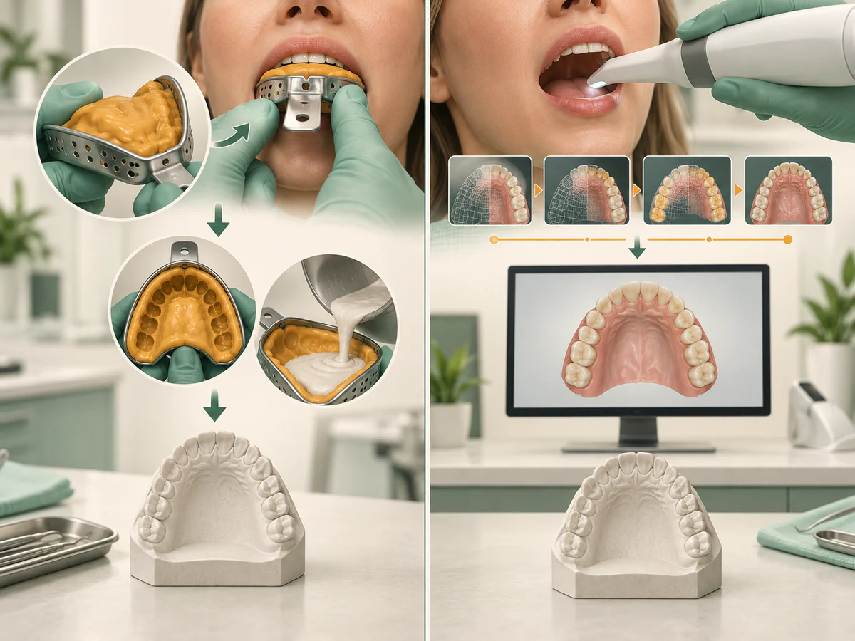
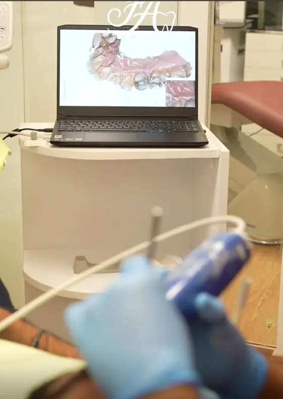
The digital impression does away with that paste. The practitioner moves the scanner camera along the teeth; the software gradually reconstructs a three-dimensional model. The resulting file can be sent directly to the laboratory or used by a computer-aided design and manufacturing chain (CAD/CAM). Removing the impression material is one of the most tangible changes for the patient, particularly for people who are sensitive to the gag reflex.
It is worth staying measured, however: digital does not automatically mean superior. Both techniques aim at the same goal, a faithful model of the arches. The choice depends on the clinical situation, and the conventional impression keeps its indications, in particular in certain cases of extensive tooth loss, discussed further on.
Comfort: what the studies say
Comfort is the benefit patients feel most clearly, and it is supported by evidence. In a crossover trial by Yuzbasioglu and colleagues (BMC Oral Health, 2014), every participant preferred the digital impression to the conventional one. Comfort scores were clearly better, particularly for the sensation of gagging and for difficulty breathing, and the total duration of the procedure was shorter (about 248 seconds versus 605 seconds for the conventional technique). One useful nuance: in that same study, the level of anxiety measured was not significantly different between the two methods. The scanner therefore improves the sensory experience of taking an impression, without removing the apprehension that comes with dental treatment.
This trend is confirmed on a larger scale. The systematic review by Siqueira and colleagues (Clinical Oral Investigations, 2021), covering 17 studies and more than 400 patients, concludes that the intraoral scanner was overall faster than the conventional impression and generally preferred by patients, whether the area scanned was a single section or a full arch, and for restorations on teeth as well as on implants. The umbrella review by Singh and colleagues (Cureus, 2025) points in the same direction: the scanner reduces procedure time and improves patient comfort.
Reducing the gag reflex is one of the most tangible contributions, since it is precisely the prolonged contact of the paste at the back of the mouth that often triggers it. Even so, no scanner makes an impression pleasant for everyone; it makes the step more tolerable for many. It is a documented improvement in comfort, not a universal promise.
Accuracy: a real advantage, but with limits worth knowing
The accuracy of an impression is described by two notions drawn from the ISO standards: trueness (how close the model obtained is to the reality of the object) and precision (the reproducibility of one recording to the next). It is their combination that defines the accuracy of an impression, and this is where the literature calls for nuance depending on the span that is scanned.
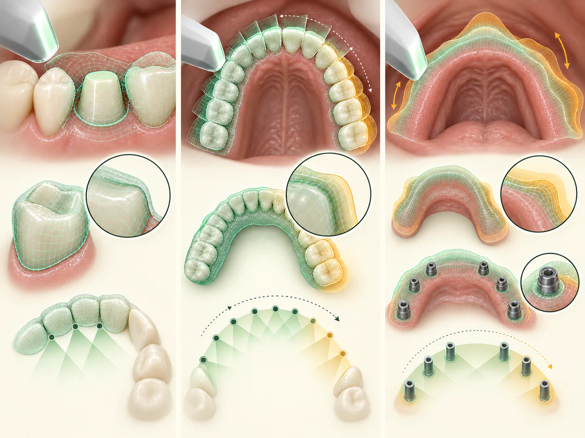
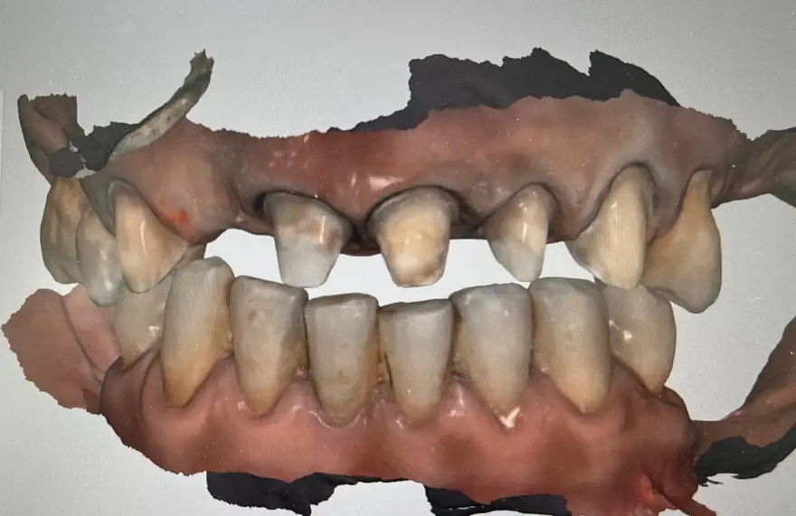
Over small spans, a single tooth or a small section, the digital impression generally reaches an accuracy comparable to that of the conventional impression, judged clinically acceptable for making well-fitting prostheses. The difficulty arises as the scanned area grows larger: stitching the images together can accumulate slight deviations over long distances.
The systematic review with meta-analysis by Pesce and colleagues (Journal of Clinical Medicine, 2024) illustrates this caution. While most of the included studies report comparable accuracy between digital and conventional impressions over the full arch, the authors describe the results as contradictory and note, in the pooled analysis, a mean difference of about 152 micrometers between the two techniques. They nonetheless conclude that, in the majority of clinical work, the accuracy of prostheses made from digital impressions remains within clinically acceptable thresholds. In other words: excellent over small spans, to be interpreted with discernment over large ones.
This is more marked still in the fully edentulous patient. The systematic review by Srivastava and colleagues (Dentistry Journal, 2023) shows that the scanner correctly reproduces well-defined anatomical landmarks, such as hard tissues covered by attached mucosa, but that the largest discrepancies appear at the peripheral borders, at the internal seal and on mobile structures or those that are hard to trace, such as the soft palate. For complex rehabilitations on several implants, the umbrella review by Singh and colleagues (Cureus, 2025) is explicit: no scanner was judged sufficiently accurate for partially edentulous arches, and conventional workflows remain preferable in multi-implant or fully edentulous situations.
| Clinical situation | Accuracy of the digital impression according to the literature |
|---|---|
| Single tooth, small section | Comparable to the conventional impression, clinically acceptable |
| Full dentate arch | Broadly comparable, but contradictory results; caution |
| Several implants, large span | Variable depending on the scanning strategy; deviations possible |
| Fully edentulous | Hard landmarks well reproduced; discrepancies at the borders and the soft palate |
This honesty about the limits takes nothing away from the value of the method: it helps to know when it is most relevant.
What is a digital impression actually used for?
The optical impression is the first link in many treatments. It feeds directly into computer-aided design, which makes it a versatile tool.
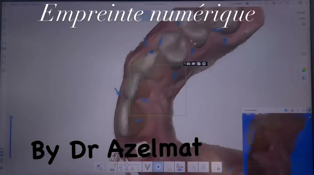
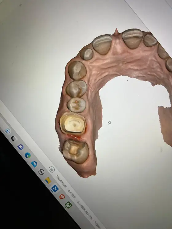
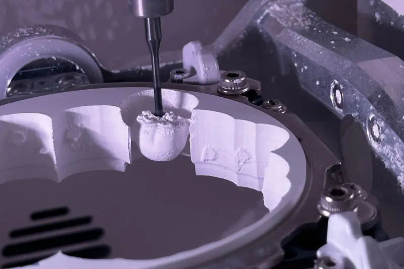
- Crowns and bridges: the file is used to design and mill the prosthesis, following on from what is described in our article on the ceramic or zirconia dental crown.
- Veneers: for these thin restorations of the front teeth, the digital impression makes esthetic communication with the laboratory easier; the subject is covered in detail in our article on ceramic dental veneers.
- Aligners and orthodontic appliances: the digital model replaces plaster casts when planning a treatment, an approach discussed in our article on orthodontics for adults in Kénitra.
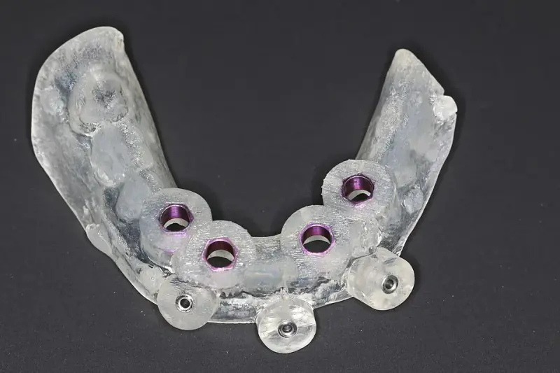
- Implant surgical guides: combined with three-dimensional imaging, the digital impression contributes to implant planning. Our guide explains dental implant placement step by step.
In these uses, the scanner does not replace clinical judgment: it provides a starting point of data, on which the design, the manufacture and the fitting then rest. The review by Ma and colleagues (International Journal of Implant Dentistry, 2023) is a reminder that, for implant impressions, accuracy varies considerably depending on the scanning strategy, with deviations reported from 27 to 360 micrometers in partially edentulous arches. That shows how much the recording technique matters, and not only the device.
The digital workflow, step by step
The scanner is part of a logical chain, from recording to prosthesis. In outline, the workflow runs as follows:
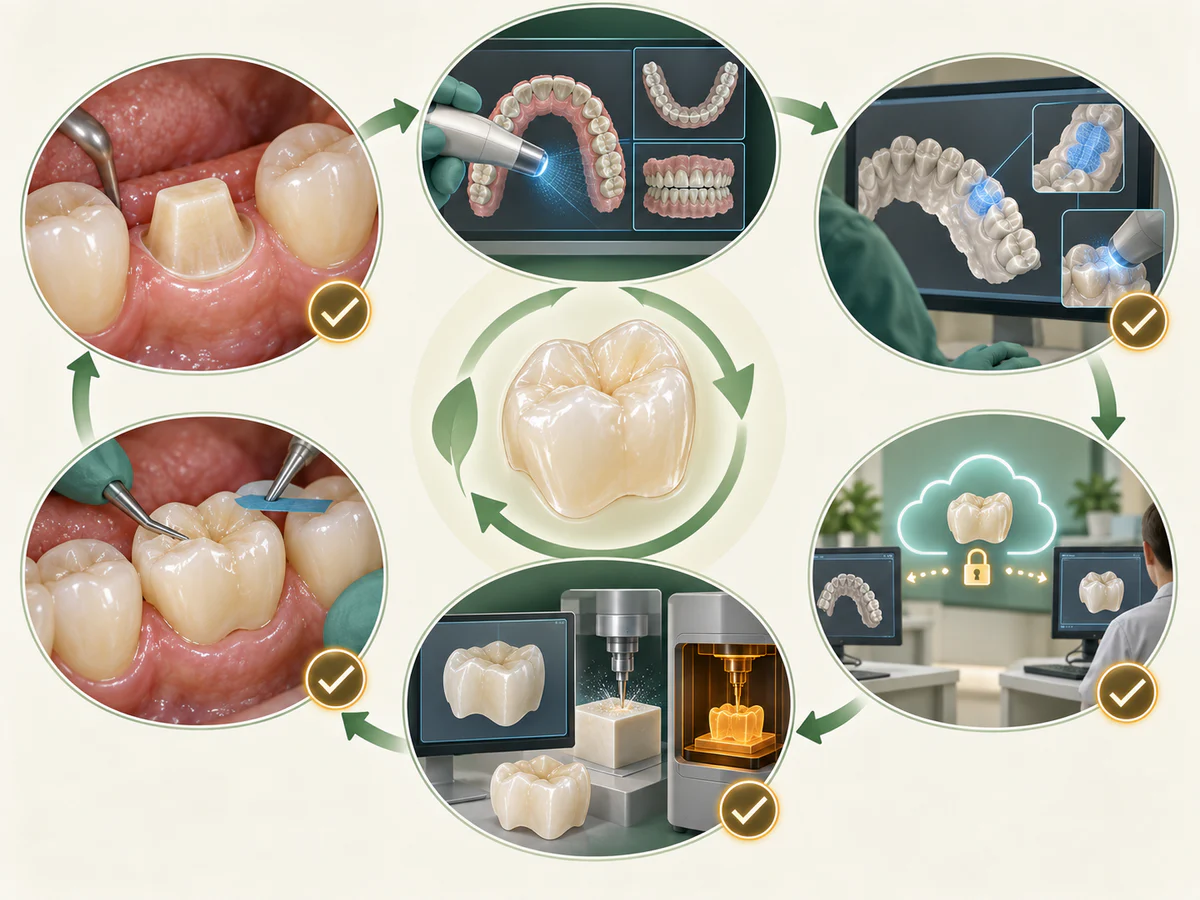
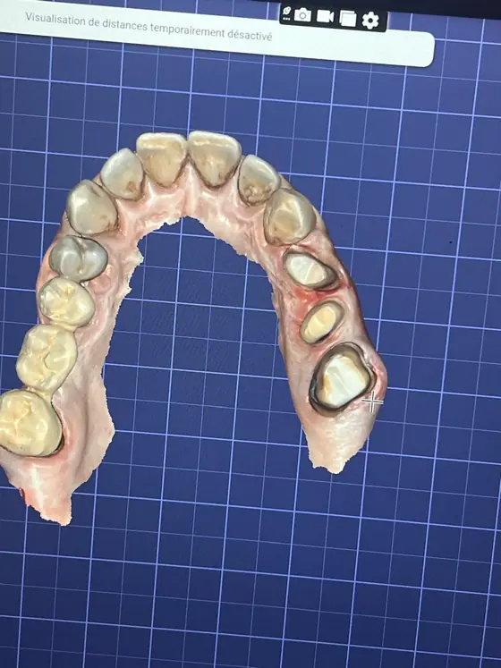
- preparation of the tooth and the gum, a step that determines how readable the margins to be recorded will be;
- optical recording of the arches and of the occlusion with the intraoral scanner;
- checking the model on screen, and immediately re-recording any incomplete areas if needed;
- transmission of the file to the laboratory or to the CAD/CAM chain;
- design and then manufacture of the prosthesis or the appliance;
- try-in in the mouth, adjustment and fitting.
One of the practical advantages of this workflow is being able to check the impression in real time: an area that was poorly recorded can be rescanned on the spot, without starting the whole procedure over. Seeing the model on screen also helps to explain the situation to the patient. This does not remove the need for rigor: a preparation margin drowned in saliva or blood, or a bleeding gum, records poorly, digitally as well as conventionally. Healthy gums remain a prerequisite, and our articles on daily dental hygiene and on bleeding gums explain why.
Digital and conventional: two techniques that coexist
The digital impression does not make the conventional impression obsolete. Both methods are tools, chosen according to the case. For a single crown, a veneer or an aligner treatment, the scanner offers greater comfort and an accuracy suited to the task. For certain fully edentulous situations or large implant rehabilitations, the data call for caution and the conventional impression, or complementary techniques, fully keep their place.
Having an intraoral scanner at the practice therefore describes a piece of equipment and a way of working, not a guarantee of results. What makes care good is the indication established after examination, the precision of the procedure and follow-up over time. The digital impression contributes to that, modestly and usefully, by making one step more comfortable and, in many cases, just as faithful.
Frequently asked questions
Is a digital impression more comfortable than a conventional impression?
Does the intraoral scanner reduce the gag reflex?
Is a digital impression as accurate as a conventional impression?
Can everything be done with a digital impression?
Is the digital impression file used to make the prosthesis?
Do you need healthy gums for a successful digital impression?
Sources
Medical references consulted for this article.
- 1Siqueira R et al., Intraoral scanning reduces procedure time and improves patient comfort in fixed prosthodontics and implant dentistry (revue systématique), Clinical Oral Investigations, 2021
- 2Yuzbasioglu E et al., Comparison of digital and conventional impression techniques: patient perception, comfort, effectiveness (essai croisé), BMC Oral Health, 2014
- 3Pesce P et al., Accuracy of Full-Arch Intraoral Scans Versus Conventional Impression (revue systématique et méta-analyse), Journal of Clinical Medicine, 2024
- 4Srivastava G et al., Accuracy of Intraoral Scanner for Recording Completely Edentulous Arches (revue systématique), Dentistry Journal, 2023
- 5Singh S et al., Accuracy and Clinical Performance of Intraoral Scanners Compared to Conventional and Extraoral Impressions (revue parapluie), Cureus, 2025
- 6Ma B et al., Accuracy of digital implant impressions obtained using intraoral scanners (revue systématique et méta-analyse, études in vivo), International Journal of Implant Dentistry, 2023
Contents
Share