Guided implant surgery involves placing a dental implant by following a surgical guide designed in advance on a computer. This guide is manufactured from two examinations: 3D imaging (the CBCT, or cone beam scan), which shows the bone volume and the position of the structures that must be protected, and a digital impression of the mouth, which captures the shape of the teeth and gums. By combining these two datasets, the ideal implant position is planned virtually, then transferred to the operating room via a guide that directs the drilling. At our practice in Kenitra, we use this approach in situations where it offers a genuine benefit: it makes it possible to place the implant in a planned position with millimeter-level precision, often without an extensive incision, for a faster, more comfortable procedure.
This article explains what guided surgery involves, how the guide is built, what the literature says about its accuracy and benefits, and above all what it does not provide. Because guided surgery is a planning and transfer tool: it helps execute a carefully considered treatment plan, but it replaces neither clinical judgment, nor bone quality, nor follow-up care, and it guarantees no outcome. Honest information means explaining both what it makes possible and where its limits lie.
What is guided implant surgery?
In so-called “freehand” implant placement, the practitioner positions the implant based on the clinical examination, radiographs, and experience, without a physical template guiding the drill. Guided surgery adds a preliminary digital planning step and a device that materializes that plan during the procedure.
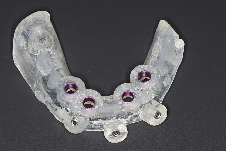
The principle unfolds in three stages. First, the implant position is planned virtually in software, by superimposing the 3D imaging and the digital impression. Next, based on this plan, a surgical guide is designed: a piece, most often made of resin, that fits over the teeth, gums, or bone and contains sleeves that direct the drill along the planned axis, depth, and position. Finally, on the day of the procedure, the guide is seated and placement follows the established plan.
There are two main families of computer-assisted techniques. Static surgery relies on a physical guide that reproduces the planned position without allowing it to be modified during the procedure. Dynamic surgery, or navigation, uses a system that tracks the instruments in real time on a screen and permits adjustments during the operation. The ITI Group 5 consensus report (Wismeijer et al., 2018) clearly distinguishes these two approaches. This article focuses mainly on static, template-guided surgery, the most widely used at our practice.
How is the surgical guide built?
The guide is not designed at random: it is the product of a digital workflow that starts with the patient and returns to the patient. Every step must be carefully controlled, because approximations add up.
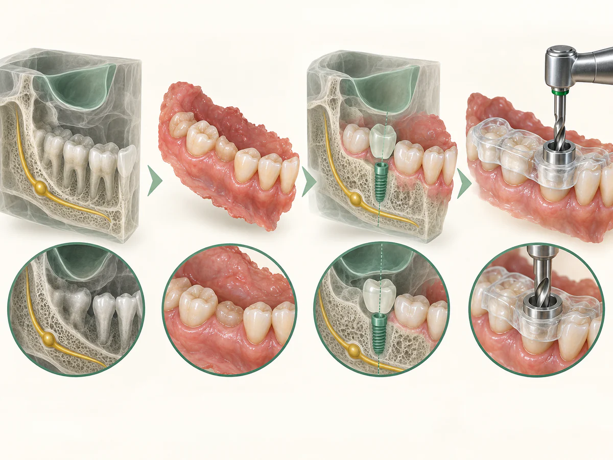
From the CBCT and the impression to the treatment plan
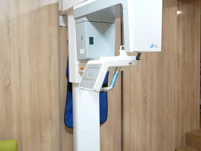
The first dataset is the CBCT (cone beam) scan, which provides a three-dimensional image of the bone and locates the structures that must not be injured, such as the inferior alveolar nerve or the maxillary sinuses. The second is the digital impression taken with an intraoral scanner, which captures the surface of the teeth and gums in fine detail. In the software, these two acquisitions are superimposed: the bone from the 3D imaging and the surface from the impression are registered onto each other to form a complete model.
It is on this model that the implant position is defined — not only according to the available bone, but also according to the future prosthesis. This is known as “prosthetically driven” planning: we start from the desired crown and work backward to determine where the implant must sit to support it properly. The ITI consensus specifically recommends aligning surface scans, including the prosthetic plan, with the 3D volumetric data to improve accuracy.
From the plan to the guide, then to the operating room
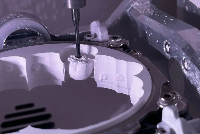
Once the position is validated, the guide is designed digitally and then manufactured, most often by 3D printing or milling. It incorporates metal sleeves that constrain the drill. On the day of the procedure, the guide is stabilized in the mouth and the drilling sequence follows the plan. When the bone and soft tissues allow it, the procedure can be performed without raising an extensive gum flap (the so-called “flapless” technique), working through the guide. Placement takes place under the usual aseptic conditions of our dedicated surgical suite, and the general steps of implant treatment are detailed in our article Dental implants in Kenitra.
What does the literature say about accuracy?
Accuracy is measured by comparing the planned implant position with its actual position after placement. Three deviations are usually assessed: at the entry point (the implant neck), at the apex (the tip), and the angular deviation (the tilt).
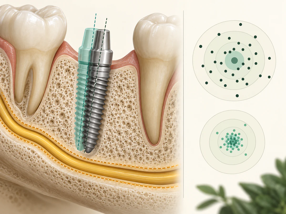
The systematic review and meta-analysis by Tahmaseb et al. (2018), published in Clinical Oral Implants Research and linked to the ITI consensus, pooled 20 studies totaling 2,238 implants in 471 patients. It reports a mean error of approximately 1.2 mm at the entry point, 1.4 mm at the apex, and 3.5° of angular deviation. The authors conclude that the accuracy of static computer-assisted surgery falls within a clinically acceptable margin in most situations, but they stress one essential point: a safety margin of at least 2 mm from at-risk structures must be maintained, because deviations always exist.
Comparison with freehand placement highlights the value of the method. The systematic review and meta-analysis by Werny et al. (2025), published in the International Journal of Implant Dentistry, found mean deviations of approximately 7.5° (angular), 1.56 mm (entry point), and 2.22 mm (apex) for freehand placement, versus approximately 2.6°, 0.72 mm, and 0.88 mm for fully guided static surgery. Placement with a full template guide is therefore considerably closer to the plan than freehand placement. The meta-analysis by Khaohoen et al. (2024), published in BMC Oral Health, places the overall deviation at around 1.1 mm at the entry point and around 1.4 mm at the apex, with an angular deviation of approximately 3.5°, and observes that robotic systems show the smallest deviations — a technology that is still not widely available.
| Approach | Mean angular deviation | Deviation at entry point | Deviation at apex |
|---|---|---|---|
| Freehand placement | ~ 7.5° | ~ 1.56 mm | ~ 2.22 mm |
| Guided, pilot drill only | ~ 5.9° | ~ 1.13 mm | ~ 1.43 mm |
| Guided, fully guided | ~ 2.6° | ~ 0.72 mm | ~ 0.88 mm |
Mean values from the meta-analysis by Werny et al. (2025); they describe trends, not a guaranteed individual result.
These figures must be read with caution: they are averages, and there is dispersion around them. A “mean” deviation of one millimeter means that some cases deviate further. That is precisely why a safety margin remains indispensable.
What concrete benefits for the patient?
Beyond measured accuracy, guided surgery aims for benefits the patient actually experiences, when it is properly indicated.
An often less invasive procedure
When the situation allows, the flapless approach avoids detaching the gum over a large area. A randomized controlled split-mouth trial published by Frizzera et al. (2021) in The International Journal of Oral & Maxillofacial Implants, comparing flapless guided surgery with conventional surgery, observed in favor of guided surgery a shorter procedure time, lower painkiller consumption, and less intra- and postoperative pain. These results point toward reduced morbidity, though they cannot be generalized to every situation.
Planning that makes the procedure safer
By materializing the axis and depth in advance, the guide helps maintain safety distances from the nerve and neighboring teeth, and position the implant according to the future prosthesis. This anticipation is particularly useful in demanding cases — limited space, proximity to an anatomical structure, or rehabilitation involving several implants.
Survival comparable to conventional surgery
The review by Tahmaseb et al. (2018), linked to the ITI consensus, reports that the few studies that tracked the survival of implants placed with a guide observed high rates at one year or more, comparable to those of conventional surgery; the data remain limited and short-term, however. The review by Joda et al. (2018) on patient-reported outcome measures (PROMs) and complications concludes, along the same lines, that the complications of static guided surgery appear negligible and comparable to conventional surgery. In other words, guided surgery does not, by itself, increase an implant’s chances of long-term survival: its contribution lies mainly in positioning accuracy and procedural comfort, not in a promise of better osseointegration.
What guided surgery does not provide
Presenting this technique honestly also means stating its limits. Guided surgery is a means, not a guarantee.
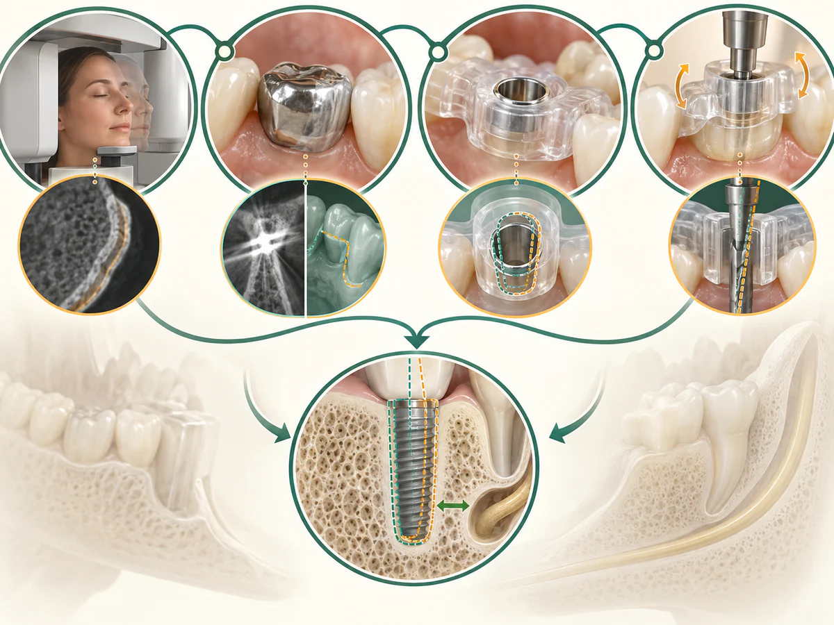
First, it is not a guarantee of results. An implant’s survival depends on the quality and volume of the bone, the condition of the gums, smoking, general health, and oral hygiene — factors the guide does not change. When bone is lacking, a bone graft or sinus lift may still be necessary before or during placement: 3D planning helps inform that decision; it does not replace it.
It does not eliminate error; it reduces it. As the analyses of error sources summarized by Tahmaseb et al. (2018) point out, deviations accumulate at every step: CBCT and impression acquisition, file superimposition, guide manufacturing, and guide stability in the mouth during drilling. Metal artifacts from old restorations can interfere with image superimposition. It is this accumulation that justifies keeping a safety margin and not treating the plan as an absolute certainty.
Finally, it is neither indicated in every situation nor indispensable. Many implants are placed successfully freehand, by an experienced practitioner, without a guide. Guided surgery requires a CBCT — and therefore X-ray exposure, justified on a case-by-case basis — plus an additional planning cost. Its value is weighed according to the complexity of the case, not as a matter of principle. The ITI consensus states no contraindication to static guided surgery, but neither does it present it as mandatory: it is one tool among others.
In which cases is it most useful?
Guided surgery comes into its own when positioning accuracy is critical: placing several implants intended to support a bridge, situations where an anatomical structure is close by, limited bone space requiring a precise axis, or the wish for a minimally invasive approach in an apprehensive patient. It is also valuable for aligning the implant with the future prosthesis from the outset, as part of a prosthetically driven treatment plan. Conversely, for a straightforward single-implant placement with abundant bone and clear landmarks, its contribution is more marginal, and the decision is made case by case, after a complete clinical examination.
In summary
Guided implant surgery transfers to the operating room a plan developed on a computer from the CBCT and the digital impression, using a guide that directs the drilling. The literature shows that it places the implant considerably closer to the planned position than freehand placement — with mean deviations on the order of a millimeter — and that it often allows a faster, less painful procedure, with implant survival comparable to conventional surgery. At our practice in Kenitra, we turn to it when the required precision justifies it. But it is neither a guarantee of success nor a routine step: deviations remain, a safety margin is still necessary, and success depends above all on the bone, the gums, and follow-up care. Guided surgery serves a carefully considered treatment plan; it does not replace it.
Frequently asked questions
Is guided implant surgery more accurate than conventional placement?
Does guided surgery guarantee the success of the implant?
Is a scan or CBCT required for guided surgery?
Is guided placement less painful than conventional surgery?
What is the difference between static guided surgery and dynamic navigation?
Do all implants need to be placed with a guide?
Sources
Medical references consulted for this article.
- 1Tahmaseb et coll. 2018, The accuracy of static computer-aided implant surgery: a systematic review and meta-analysis, Clinical Oral Implants Research (PubMed)
- 2Werny et coll. 2025, Freehand vs. computer-aided implant surgery: a systematic review and meta-analysis — part 1: accuracy of planned and placed implant position, International Journal of Implant Dentistry (PMC)
- 3Khaohoen et coll. 2024, Accuracy of implant placement with computer-aided static, dynamic, and robot-assisted surgery: a systematic review and meta-analysis of clinical trials, BMC Oral Health (PMC)
- 4Frizzera et coll. 2021, Flapless Guided Implant Surgeries Compared with Conventional Surgeries Performed by Nonexperienced Individuals: Randomized and Controlled Split-Mouth Clinical Trial, Int J Oral Maxillofac Implants (PubMed)
- 5Joda et coll. 2018, Static computer-aided implant surgery (s-CAIS) analysing patient-reported outcome measures (PROMs), economics and surgical complications: a systematic review, Clinical Oral Implants Research (PubMed)
- 6Wismeijer et coll. 2018, Group 5 ITI Consensus Report: Digital technologies, Clinical Oral Implants Research (Wiley)
- 7ITI Academy, Consensus statements on Static Computer-Aided Implant Surgery (s-CAIS): accuracy, PROMs and complications
Contents
Share