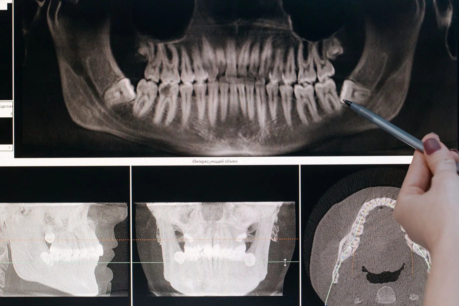
A wisdom tooth is not removed as a matter of principle. The decision to extract rests on the presence of a symptom and/or a pathology linked to that tooth, and not on its position alone. This is the guiding principle set out by the HAS in its 2019 recommendation on the removal of third molars: impaction and ectopia — that is, the fact that a tooth is impacted, tilted or poorly positioned — are not in themselves criteria for a treatment decision. The UK’s NICE takes the same view and recommends abandoning the preventive extraction of impacted wisdom teeth that show no pathology. In other words, a wisdom tooth that is asymptomatic and shows no sign of disease can be monitored rather than extracted.
This position runs counter to a still widely circulated idea, according to which most wisdom teeth will eventually cause problems and should be removed as a precaution. There is no reliable figure supporting such a claim, and the decision is made case by case. As a dental surgeon in Kénitra, I frequently see people who are worried about their wisdom teeth. This article distinguishes genuine indications for extraction from situations that call for simple monitoring, and explains the role of imaging and the follow-up of the recovery period.
When should a wisdom tooth be extracted?
Wisdom teeth, or third molars, generally appear between roughly 17 and 25 years of age. Not all of them cause problems. Many erupt normally and remain functional or asymptomatic. The useful question is therefore not “should they be removed?” but “is this particular tooth causing a symptom or a pathology?”.
The recognized indications
The HAS sets out a list of situations in which removal is recommended. These notably include:
- decay that is symptomatic and cannot be durably restored;
- symptomatic pulpal or periapical disease that cannot be treated;
- pericoronitis that is recurrent and/or resistant to conservative treatment;
- cellulitis, an abscess or osteomyelitis related to the tooth;
- a cyst or a tumor of the follicular sac;
- internal or external resorption of the adjacent tooth;
- periodontal disease related to the third molar.
NICE lists very similar indications, which gives these criteria international consistency. What all these situations have in common is that they correspond to a confirmed symptom or to an objectively documented pathology, not simply to an unfavorable position.
The particular case of pericoronitis
Pericoronitis is inflammation of the gum tissue that partly covers an erupting wisdom tooth. It is one of the most common reasons for consultation. An isolated episode does not automatically justify extraction: it can be treated conservatively. According to the HAS, it is when this inflammation becomes recurrent and/or resistant to treatment that it becomes an indication for removal. The distinction matters, because it avoids hasty extractions after a first episode that can often be brought under control.
When pericoronitis is accompanied by more marked signs — swelling, intense pain, difficulty opening the mouth or fever — prompt care is needed. Our guide to what to do right away in a dental emergency sets out the situations that call for a rapid consultation.
When should you monitor rather than extract?
This is where the gap is widest with any message that pushes for systematic extraction. In the absence of symptoms, in a healthy adult over 30, the HAS does not recommend the systematic removal of a third molar. The position of the tooth, even impacted or horizontal, is not on its own enough to decide to take it out.
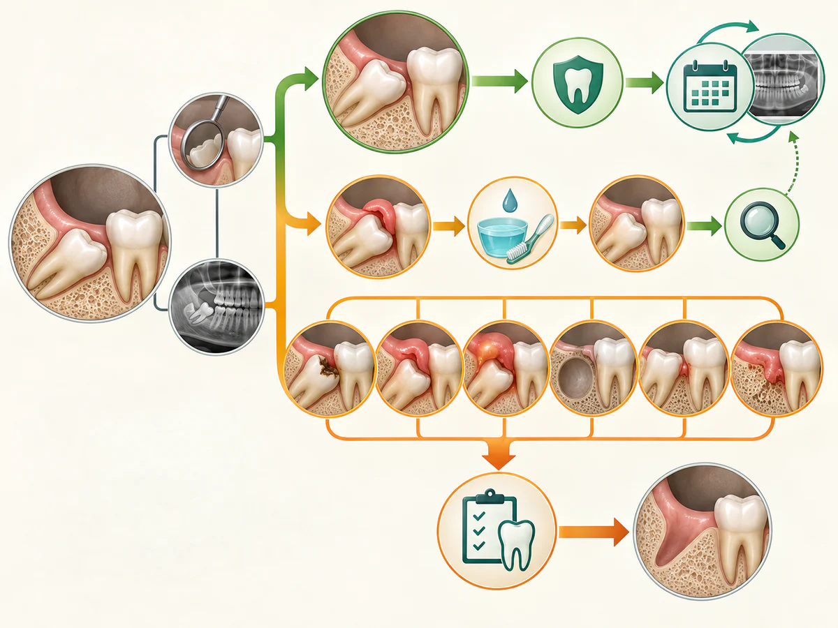
The 2020 Cochrane review, which compared extraction with retention of asymptomatic, disease-free impacted wisdom teeth, concludes that the available data are of low to very low certainty. They do not make it possible to state that these teeth must be removed. Retention could be associated with an increased risk of periodontal problems on the neighboring second molar, but that finding is itself of very low certainty. In this context of uncertainty, the reasonable approach is a decision shared with the patient and, if the tooth is kept, periodic clinical and radiographic follow-up.
The orthodontic argument does not hold up
One justification comes up often: wisdom teeth would need to be removed to keep the other teeth from crowding, or to protect the result of orthodontic treatment. This idea is not supported. The HAS states clearly that removing a third molar is not recommended in order to prevent anterior crowding. The systematic review by Lyros et al. (Dentistry Journal 2023) finds no proven link between the lower wisdom teeth and the relapse of incisor crowding after orthodontic treatment, and finds insufficient data to advise their preventive extraction in the name of occlusal stability. Extracting for this reason is therefore not justified.
What imaging work-up before the extraction?
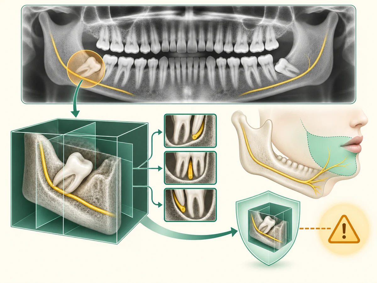
Imaging serves to assess the position of the tooth, its roots and its anatomical relationships, in particular with the inferior alveolar nerve for the lower wisdom teeth.
The panoramic X-ray as first-line imaging
According to the HAS, the first-line radiological work-up is the dental panoramic X-ray. It gives an overall view of both arches and is sufficient in the majority of cases to plan an extraction. Turning straight to three-dimensional imaging for every wisdom tooth is not justified.
When is the Cone Beam useful?
The Cone Beam, or cone beam computed tomography, is reserved for situations in which the lower wisdom tooth appears to be in close relationship with the canal of the inferior alveolar nerve on the panoramic X-ray. It then provides a precise three-dimensional reading of the relationship between the roots and the canal, useful for surgical planning and for informing the patient. At the practice, in Kénitra, we have a Durr Dental Cone Beam, which we reserve for those cases where a detailed analysis of the nerve’s course genuinely changes the surgical strategy.
One point deserves to be stated without ambiguity, because it is often oversold. The Cone Beam does not eliminate the nerve risk. The meta-analysis by Del Lhano et al. (Dentomaxillofacial Radiology, 2020) compared the panoramic X-ray with the Cone Beam and did not demonstrate any significant reduction in inferior alveolar nerve paresthesia in favor of the Cone Beam (relative risk 1.23; 95% confidence interval 0.75 to 2.02; p equal to 0.43). Three-dimensional imaging improves mapping and decision-making, but it has not been shown to lower the actual rate of nerve injury. Promising planning that would avoid any nerve damage would be inaccurate.
How does the procedure take place?
The extraction is carried out under local anesthesia to control sharp pain during the procedure. Pressure, pulling or vibration may still be felt; tell the clinician immediately if you feel pain. Depending on the position of the tooth, the operation may be straightforward or may require a surgical approach: an incision in the gum, uncovering of the bone, and sometimes sectioning of the tooth into several fragments so that it can be removed without straining the neighboring structures.
Things have to be stated accurately: the anesthetic is used to control pain during the procedure, but pressure or movement may still be felt and the operation carries real risks. Pain and swelling after the operation are expected, and complications do exist. It is precisely because these risks are real that they are explained and managed before the procedure. Our surgical facilities, which include a dedicated operating room, piezosurgery for a more selective bone cut and Choukroun PRF prepared from the patient’s own blood, are intended to carry out these extractions under good conditions, without for all that removing the risks inherent in any surgery.
What are the possible after-effects and complications?
Telling normal after-effects apart from complications helps you know when to be concerned.
Normal after-effects
Swelling and discomfort are generally at their worst in the first few days, then subside. Soft-tissue healing usually takes about one to two weeks, but these timeframes vary from one person to another and are not a guarantee. It is not abnormal to have slightly limited mouth opening and passing sensitivity during this period.
Injury to the inferior alveolar nerve
This is the complication specific to the lower wisdom teeth that warrants the most caution. Injury to the inferior alveolar nerve leads to reduced or lost sensation in the lower lip and the chin on the affected side, and sometimes to abnormal sensations. According to Del Lhano et al. (2020), the risk of temporary injury associated with the extraction of third molars lies between 0.4% and 6%, and the risk of permanent injury, meaning injury that persists beyond six months, is below 1%. Most often, then, the disturbance is temporary, but permanent damage remains possible, albeit rare. The lingual nerve may also be affected.
Dry socket
Dry socket corresponds to the loss or the absence of the blood clot that should normally fill the socket after the extraction. It shows itself as pain that appears or intensifies a few days after the procedure. It is more frequent after the removal of the lower wisdom teeth than after a simple extraction. Smoking is a major risk factor: according to the systematic review by Kuśnierek et al. (Dentistry Journal 2022), it multiplies the risk roughly threefold, with a frequency of around 13% in smokers compared with around 4% in non-smokers. Other factors play a part, such as a difficult extraction, a pre-existing infection or the use of oral contraceptives. Smoking is therefore an important contributor, but dry socket remains multifactorial in origin.
| Element | Normal after-effect | Warning sign |
|---|---|---|
| Pain | Present, then decreasing over the first few days | Pain that returns or worsens after 2 to 4 days |
| Swelling | At its worst at the start, then subsiding | Swelling that increases along with fever |
| Sensation in the lip or the chin | Normal | Persistent numbness on the operated side |
| Bleeding | Slight initial oozing | Heavy bleeding that does not stop |
How can you recover well after the extraction?
The priority in the first few hours is to protect the blood clot, which is essential to good healing. The UFSBD advice sheet gives clear recommendations:
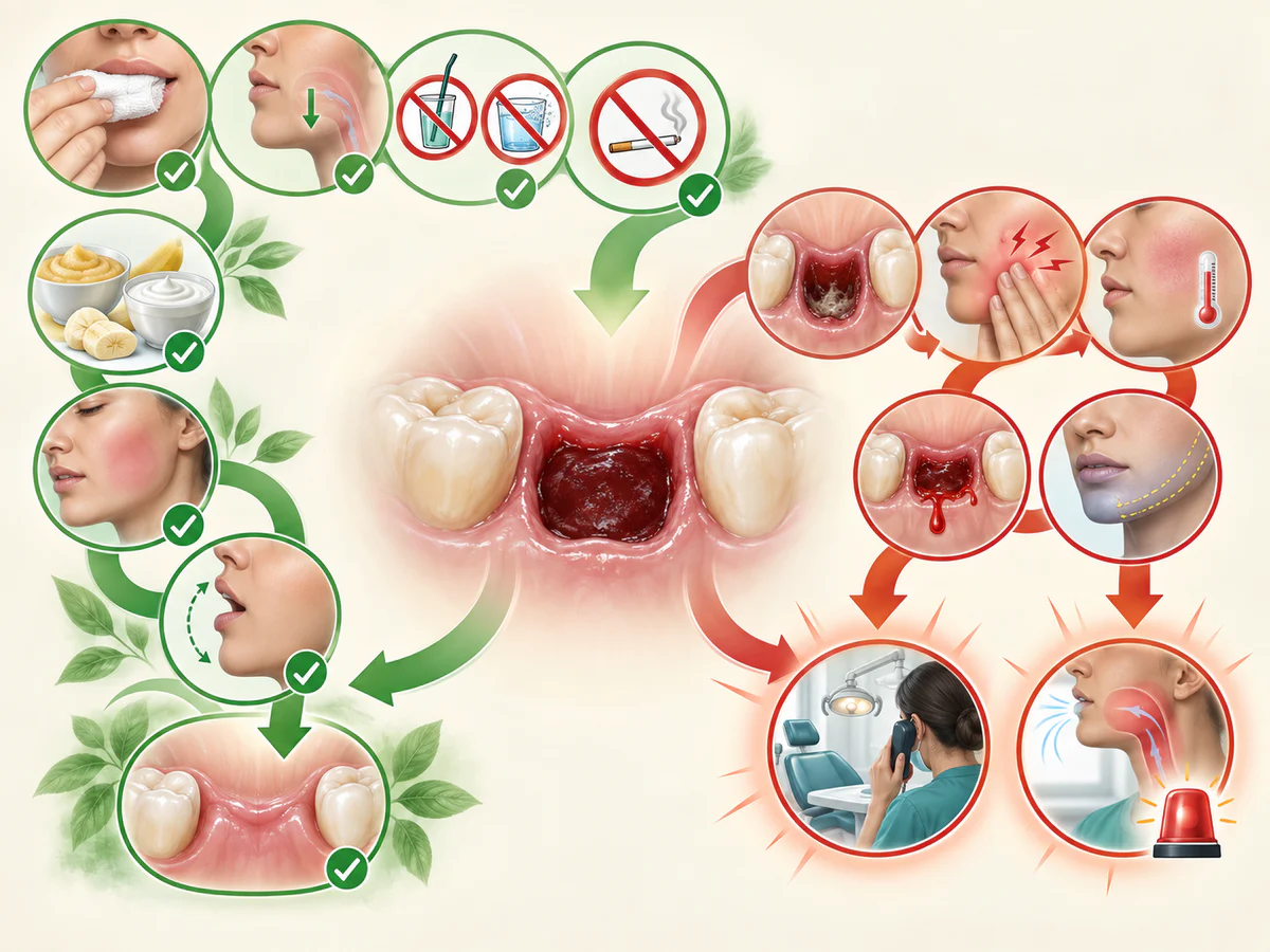
- bite on the gauze pad for about thirty minutes after the procedure, to encourage the clot to form;
- swallow your saliva normally and do not spit, so as to avoid causing bleeding;
- do not rinse the mouth vigorously, and do not use a straw;
- do not smoke, as smoking delays healing and encourages dry socket;
- just after the extraction, for as long as the anesthetic is working, do not eat and do not drink alcohol, so as to avoid injuring yourself without feeling it.
As for antibiotics, they are not a routine treatment after an extraction. They are reserved for situations of confirmed infection or general signs, and prescribing them is a medical decision made case by case.
And afterwards, does the tooth need to be replaced?
In the vast majority of cases, an extracted wisdom tooth does not need to be replaced, because it does not take part in the bite in the same way as the other molars. Replacement mainly concerns the molars further forward, whose absence can affect chewing and the balance of the dental arches.
When another tooth does need to be replaced, several options exist depending on the clinical situation. Our article comparing an implant, a bridge or dentures details the criteria for choosing. And when the bone volume is insufficient to place an implant, reconstruction solutions exist, presented in our guide to dental implants when there is not enough bone.
In summary
Removing a wisdom tooth is neither a procedure to be taken lightly nor an inevitable rite of passage. According to the HAS and NICE, it is decided on the basis of a symptom and/or a pathology, never on the position of the tooth alone: an impacted tooth that is asymptomatic and shows no sign of disease can be monitored. The panoramic X-ray remains the first-line imaging, with the Cone Beam reserved for close relationships with the inferior alveolar nerve, without it reducing the nerve risk for all that. The procedure is well controlled but carries real risks, in particular nerve injury and dry socket, whose prevention relies in part on stopping smoking and protecting the clot. In the event of recurrent pericoronitis, pain or swelling, an examination makes it possible to pinpoint the indication precisely and to avoid both under-treatment and unnecessary extraction.
How the procedure runs at the practice, the expected recovery and the fees are set out on the wisdom tooth removal in Kénitra page.
Frequently asked questions
Should an impacted wisdom tooth that does not hurt be removed?
Is having a wisdom tooth extracted painful?
How long does the recovery last after a wisdom tooth extraction?
What is dry socket and how can it be avoided?
Can a wisdom tooth shift my other teeth?
Is the Cone Beam always necessary before the extraction?
Sources
Medical references consulted for this article.
- 1HAS, Avulsion des 3es molaires : indications, techniques et modalités, recommandation de bonne pratique, mai 2019
- 2NICE, Guidance on the extraction of wisdom teeth (TA1), chapitre 1 Guidance
- 3Cochrane Oral Health, Ghaeminia et coll., Surgical removal versus retention for asymptomatic disease-free impacted wisdom teeth, CDSR 2020, CD003879
- 4Del Lhano NC et coll., Panoramic versus CBCT to reduce IAN paresthesia after third molar extractions, Dentomaxillofac Radiol 2020;49(4)
- 5Lyros I et coll., The Effect of Third Molars on Mandibular Anterior Crowding Relapse: A Systematic Review, Dent J 2023;11(5):131
- 6Kuśnierek W et coll., Smoking as a Risk Factor for Dry Socket: A Systematic Review, Dent J 2022;10(7):121
- 7UFSBD, Conseils post-opératoires après une extraction dentaire, fiche conseil
Contents
Share