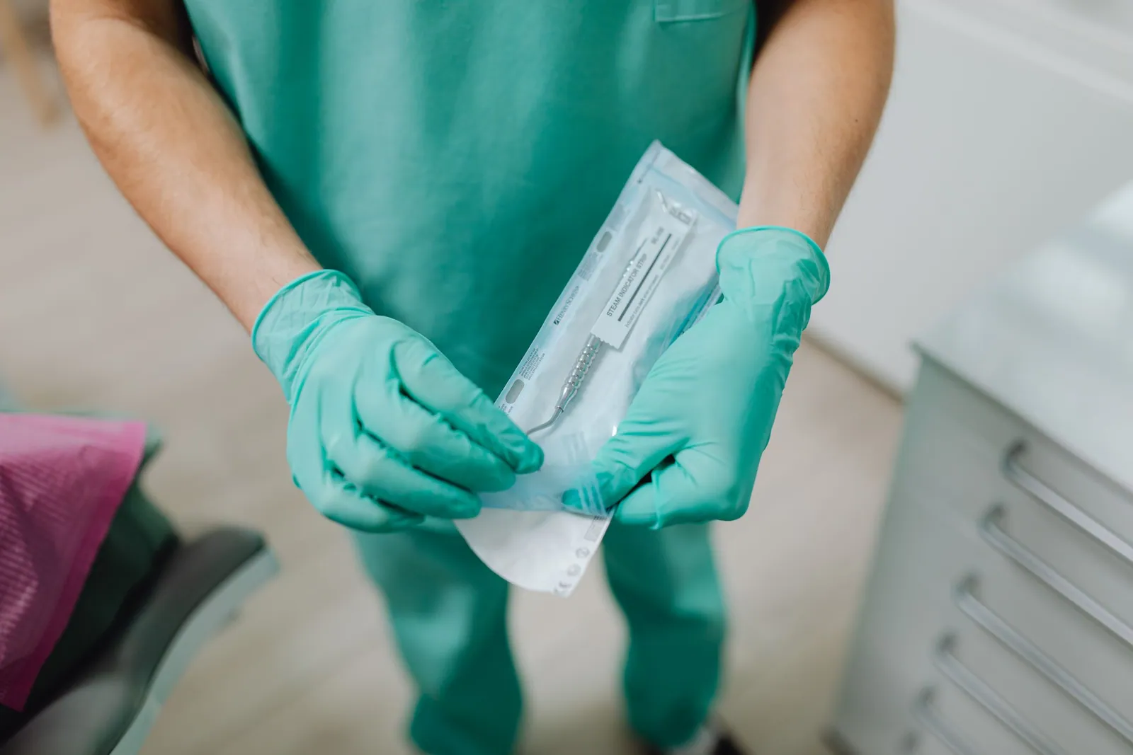
Placing an implant, performing a bone graft, or extracting an impacted tooth are not routine dental treatments: they are surgical procedures that open direct access to the bone and the deep tissues. That is why they are not carried out in an ordinary treatment chair, but in an environment prepared to keep the entry of microorganisms to an absolute minimum. At the practice in Kenitra, these procedures take place in a dedicated surgical suite — a space reserved for surgery, organized around one simple rule: everything that comes into contact with the operative site must be sterile, and everything moving around it must be controlled. This article describes, without unnecessary jargon, how this asepsis chain works, which guidelines it draws on, and why it matters for the success of an implant.
It must be said plainly and from the outset: no hygiene measure, however rigorous, completely eliminates the risk of infection. Asepsis protocols reduce this risk; they do not cancel it out. It is precisely because zero risk does not exist that these procedures are so tightly governed, repeated, and verified. Presenting a surgical suite as an absolute guarantee against infection would be overpromising; presenting it as a set of serious precautions that tip the odds in your favor is accurate.
Why a dedicated environment for surgery?
The skin and mucous membranes form a natural barrier against microorganisms. Any surgical procedure breaches this barrier: it creates a point of entry into tissues that are normally protected. The mouth, moreover, is permanently home to an abundant microbial flora. The challenge of oral surgery is therefore twofold: not to add germs from outside, and to limit the passage of germs already present into the surgical wound.
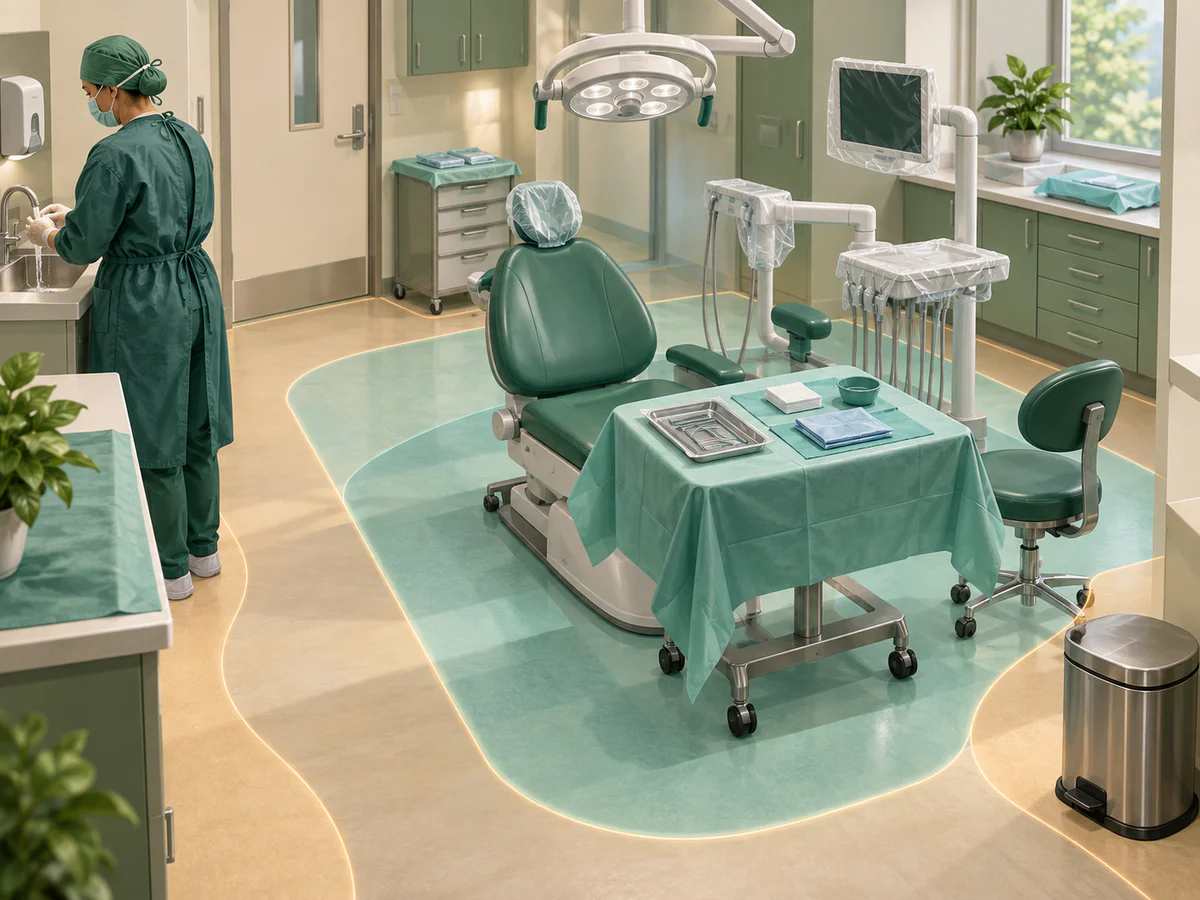
A dedicated environment answers this logic. It separates what is clean and sterile from what is not, organizes the movement of equipment and people, and brings together the conditions that favor a safe procedure. This concern is not unique to dentistry: it lies at the very heart of international surgical guidelines.
The World Health Organization (WHO), in its Global Guidelines for the Prevention of Surgical Site Infection (2nd edition, 2018), brings together the evidence-based measures to apply before, during, and after surgery to reduce these infections. The guiding principle is consistent: prepare the patient, prepare the team, prepare the instruments and the environment. Implant surgery, though on a small scale, follows this same philosophy.
The implant is placed in the surgical suite under local anesthesia, as described in our article on how a dental implant is placed, step by step.
The asepsis chain, link by link
Asepsis is not a single action but a chain: it takes only one link to fail for the whole to lose its value. It can be described as several stages that follow one another around the procedure.
Hand hygiene and preparing the team
The first link is also the best documented: hand hygiene. The WHO Guidelines on Hand Hygiene in Health Care (2009) popularized the framework of the “Five Moments for Hand Hygiene,” which pinpoints the key moments when the caregiver must disinfect their hands to interrupt transmission. For a surgical procedure, the document also describes a specific surgical hand preparation — more thorough than routine washing — carried out either with an antimicrobial soap or with a suitable alcohol-based hand rub.
Added to this are surgical attire, sterile gloves, a mask, and eye protection. This equipment protects the patient as well as the practitioner, and contributes to the barrier between the team and the operative site.
The sterile field and site preparation
A sterile field is set up around the surgical area: textile or single-use drapes delineate a clean surface on which only sterile items will be placed. The site itself, in and around the mouth, is prepared with antisepsis. The aim is not to achieve a sterility that is impossible inside an oral cavity, but to lower the microbial load at the time and place of the procedure.
The separation between the sterile zone and the rest of the room structures all movement during the procedure. It is this spatial discipline, more than any particular product, that gives the chain its strength.
Instrument sterilization
The instruments that penetrate tissues are, by definition, those that carry the greatest risk of infection. The US Centers for Disease Control and Prevention (CDC) classify devices into three categories according to their use: “critical” instruments, which penetrate soft tissue or bone, such as surgical instruments; “semi-critical” instruments, which touch mucous membranes; and “non-critical” instruments, which touch only intact skin. The CDC are explicit: critical instruments must always be heat-sterilized.
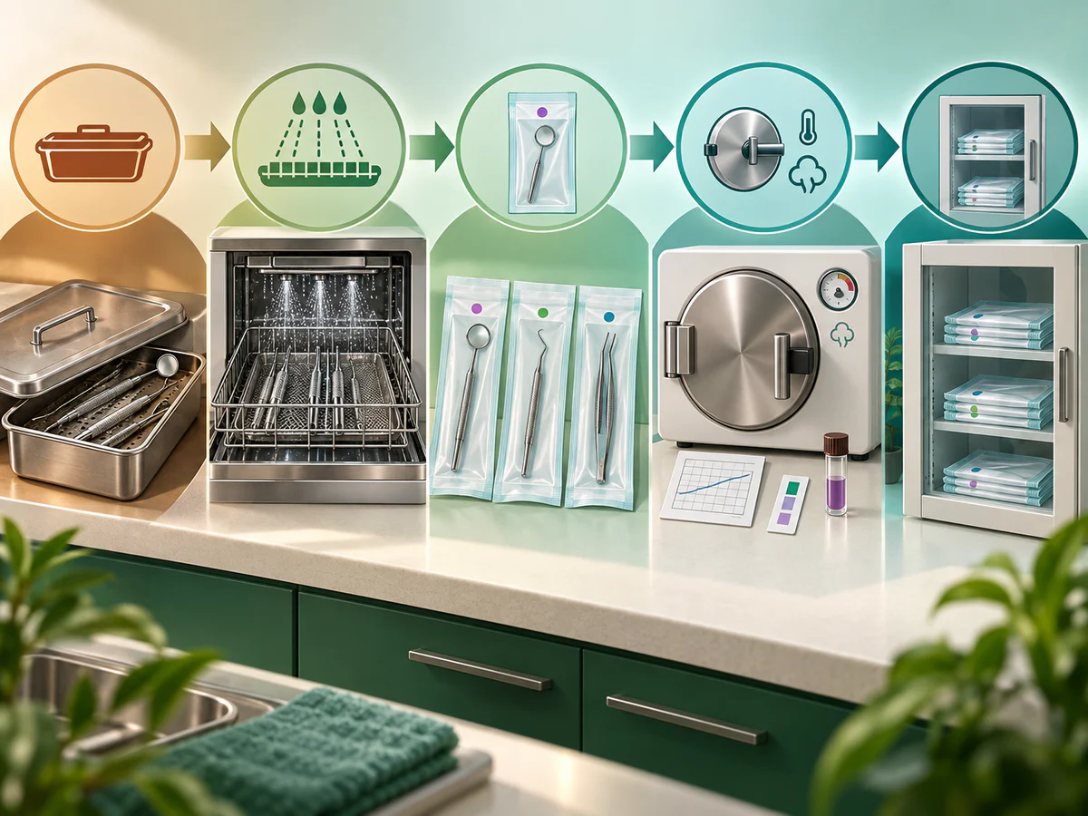
Sterilization is not a single action but a sequence of steps to follow in order, every time: cleaning, packaging, autoclaving, monitoring, then protected storage. In France, the professional recommendations relayed by the French Dental Association (Association dentaire française, ADF) and standard practice call for, for medical devices in dental surgery, a steam sterilization cycle of the type suited to porous and hollow loads, at 134 °C. The layout of the premises follows the “forward flow” principle (marche en avant): equipment moves from dirty to clean without ever going back, which prevents recontamination.
Verification: proving, not assuming
An essential link, often invisible to the patient, is the monitoring of sterilization. You do not assume that an instrument is sterile: you verify it. The CDC recommend combining three types of sterilizer monitoring.
| Type of monitoring | What it checks | Frequency recommended by the CDC |
|---|---|---|
| Mechanical | Cycle temperature, pressure, and duration reached | Every cycle |
| Chemical | Penetration of the sterilizing agent (color change of the indicator) | In every pouch |
| Biological (spore test) | Effective destruction of highly resistant spores | At least once a week |
Biological monitoring, or the spore test, is the most conclusive: it confirms that the cycle has actually destroyed highly resistant microorganisms. The CDC specify a point that is directly useful in implantology: for any load containing an implantable device, a spore test must be performed, and the implant must not be used before a negative result is obtained. This is an additional requirement that applies specifically to dental implants.
What the guidelines say about surgical safety
Beyond the instruments, the safety of a surgical procedure also depends on how the team is organized and on the checks carried out before the procedure. This is the purpose of the WHO Surgical Safety Checklist, developed as part of the “Safe Surgery Saves Lives” program. This checklist, simple and quick, was associated with a reduction in complications and mortality of more than 30% in the initial studies.
Two honest caveats accompany this figure. First, it comes from the context of major hospital surgery and does not transfer as such to office-based implant surgery; it illustrates a rationale, not a guaranteed outcome for any given patient. Second, the WHO itself points out that a significant proportion of surgery-related harm remains preventable, which implies, by extension, that complications still occur despite the best precautions. Surgical safety is a set of measures that reduce risk and that call for rigor and consistency.
In France, the reference framework for the practice is the guideline from the Haute Autorité de santé (HAS) entitled “Hygiène et prévention du risque infectieux en cabinet médical ou paramédical” (Hygiene and prevention of infection risk in medical and paramedical practices, 2007). It details standard precautions, the reprocessing of medical devices, the organization and upkeep of the premises, and the hygiene requirements for professionals. It is the foundation that, in practice, structures the asepsis chain described above.
Asepsis and the prevention of peri-implantitis
The link between asepsis and the success of an implant is not limited to the day of surgery. An implant may integrate perfectly, then later develop a disease around it: peri-implantitis, an inflammation associated with progressive bone loss.
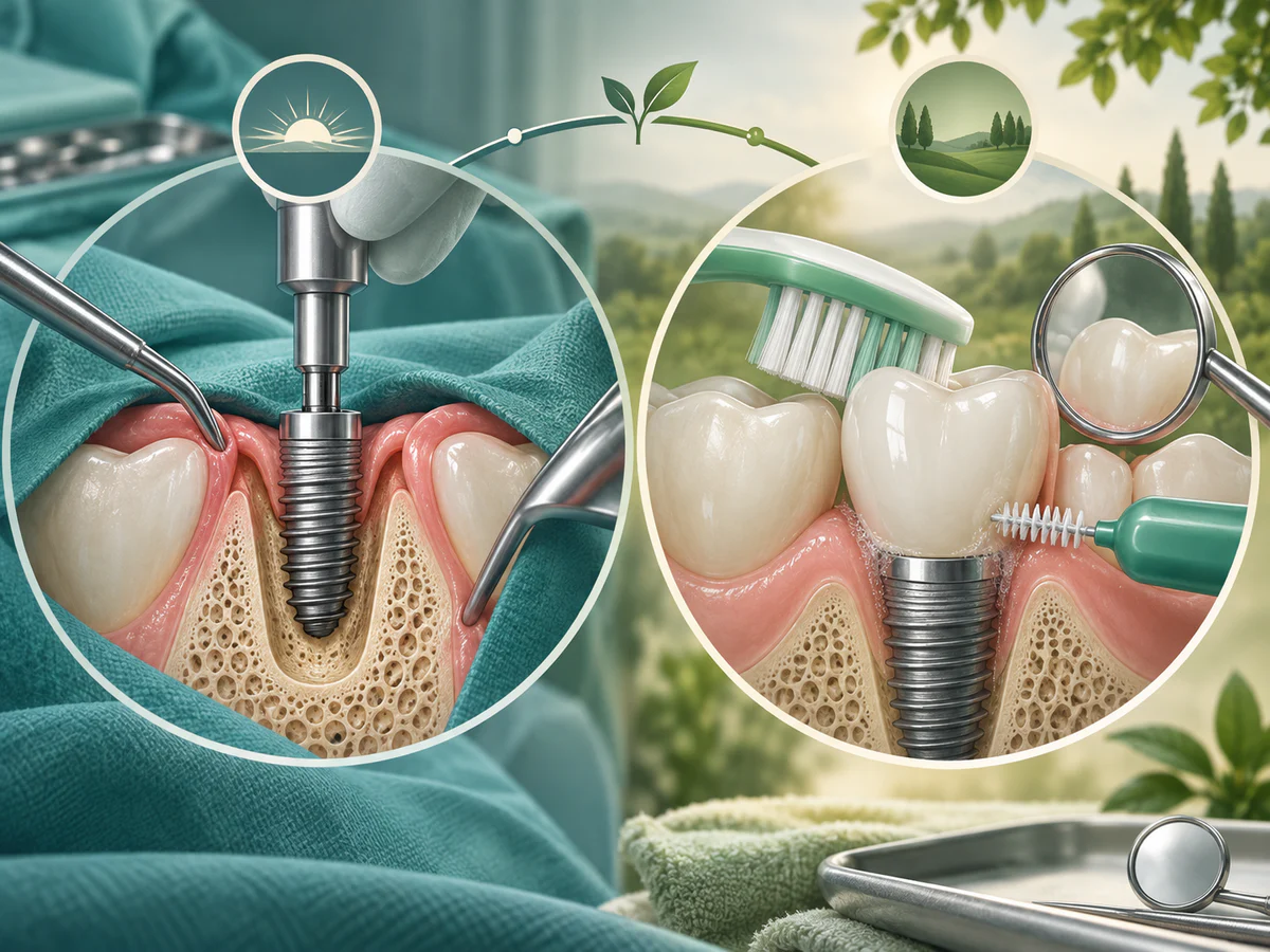
Here, we need to be precise so as not to conflate things. The main cause of peri-implantitis is not a lapse in asepsis in the surgical suite, but the accumulation of a plaque biofilm around the implant over time. The recommendations of the European Federation of Periodontology (EFP), published as an S3-level guideline (Herrera et al., 2023, Journal of Clinical Periodontology), identify this biofilm as the primary etiological factor in peri-implant diseases, and stress that prevention begins as early as implant planning, continues through surgery and loading, and then throughout the life of the implant.
Asepsis in the surgical suite and the prevention of peri-implantitis are therefore two complementary phases of the same requirement. The first aims to prevent an infection from compromising initial healing and osseointegration. The second — maintenance — aims to prevent a chronic infection from taking hold years later. An article on the maintenance of peri-implant tissues in general practice (British Dental Journal, 2024), drawing on the EFP guideline, points out that plaque control by the patient and regular professional supportive care are decisive in preventing or delaying these diseases.
In other words, the safety of an implant is determined at two moments: a controlled surgical environment at the start, then rigorous hygiene and follow-up over time. The warning signs of a late complication are described in our article on implant failure and peri-implantitis.
The special case of bone grafts and reconstructions
Bone reconstruction procedures, such as a graft before implant placement, clearly illustrate the importance of a dedicated environment. They often involve filler materials, sometimes a membrane, and open a site that will have to heal beneath the gum. Any contamination of this site can compromise the take of the graft.
That is why these procedures fall squarely within the surgical asepsis chain: sterilized and monitored instruments, a sterile field, preparation of the team and the site. Here again, these precautions reduce the risk of infection-related failure without eliminating it, because healing also depends on patient-specific factors, such as smoking or how well diabetes is controlled. The steps and conditions of a bone reconstruction are detailed in our article on bone grafting before an implant.
What the patient can observe and expect
From the patient’s point of view, several concrete signs attest to an applied asepsis chain: instruments presented in sealed pouches and opened in front of them, sterile gloves put on at the last moment, a drape placed around the surgical area, and a room prepared for the procedure.
It is just as important to understand what these measures do not promise. They do not guarantee healing free of any aftereffects, nor the complete absence of complications. They create the most favorable conditions for a safe procedure, which is a different thing. Following the post-operative instructions, daily hygiene, and follow-up all contribute, for their part, to the rest of the journey.
In summary
Implant surgery takes place in a dedicated surgical suite because it breaches the tissue barrier and requires a controlled environment. The asepsis chain that governs it rests on specific links: hand hygiene and team preparation, a sterile field and site antisepsis, heat sterilization of instruments, and documented verification of that sterilization, including a spore test before any use of an implant. These practices draw on solid references: the WHO Global Guidelines for the Prevention of Surgical Site Infection and on Hand Hygiene, the WHO Surgical Safety Checklist, the CDC classifications, the HAS guideline for the practice, and, for the prevention of peri-implantitis, the EFP guideline. All converge on the same idea, stated plainly: these protocols genuinely reduce the risk of infection, but never eliminate it. It is this cautious, verifiable, and consistent framework that serves the patient’s safety, from the day of the procedure through to long-term follow-up.
Frequently asked questions
Why isn't an implant placed in an ordinary treatment chair?
Does a surgical suite guarantee the absence of infection?
How do we know the instruments are truly sterile?
What are the sterilization chain and the "forward flow" principle?
Does asepsis in the surgical suite protect against peri-implantitis?
Which guidelines govern hygiene in dental surgery?
Sources
Medical references consulted for this article.
- 1OMS, Global Guidelines for the Prevention of Surgical Site Infection, 2e éd. 2018 (NCBI Bookshelf)
- 2OMS, WHO Guidelines on Hand Hygiene in Health Care, 2009 (NCBI Bookshelf)
- 3OMS, Safe Surgery / Surgical Safety Checklist (Une chirurgie plus sûre pour épargner des vies)
- 4CDC, Sterilization and Disinfection in Dental Settings (classification des instruments)
- 5CDC, Best Practices for Sterilization Monitoring in Dental Settings (contrôle mécanique, chimique, biologique)
- 6HAS, Hygiène et prévention du risque infectieux en cabinet médical ou paramédical, 2007
- 7Herrera et coll. 2023, EFP S3 guideline, prévention et traitement des maladies péri-implantaires, J Clin Periodontol
- 8Maintenance of peri-implant health in general dental practice, British Dental Journal 2024 (PMC11126374)
Contents
Share