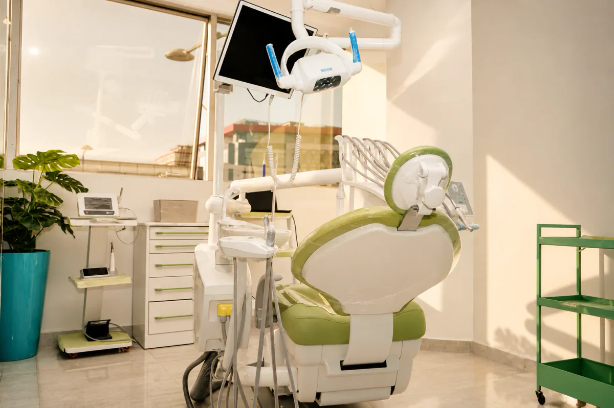
The intraoral camera is a small, pen-sized camera that the practitioner moves around your mouth to display, live and in close-up, the condition of your teeth and gums on a screen. Where you previously had to rely on a description, you can now see for yourself the crack, the tartar lodged beneath the gumline, or the old, cracked filling. This article explains, in general terms and independently of any particular piece of equipment, what this tool is for, how it changes the relationship between patient and dentist, and above all what it does not do.
Its main value is not to make a diagnosis in your place: it is a visualization and communication tool. By showing you what the practitioner observes, it helps you understand the situation, ask specific questions, and make an informed decision. The literature confirms this benefit for understanding and motivation, without making it a diagnostic instrument superior to the clinical examination. It is a useful complement, not a replacement.
What Is an Intraoral Camera?
It is an optical sensor fitted with a light source and connected to a screen. The practitioner moves it along the teeth and gums; the highly magnified image is displayed in real time. Depending on the model, the camera can freeze a photo, record a short clip, or store images in the patient’s file to compare the situation from one visit to the next.
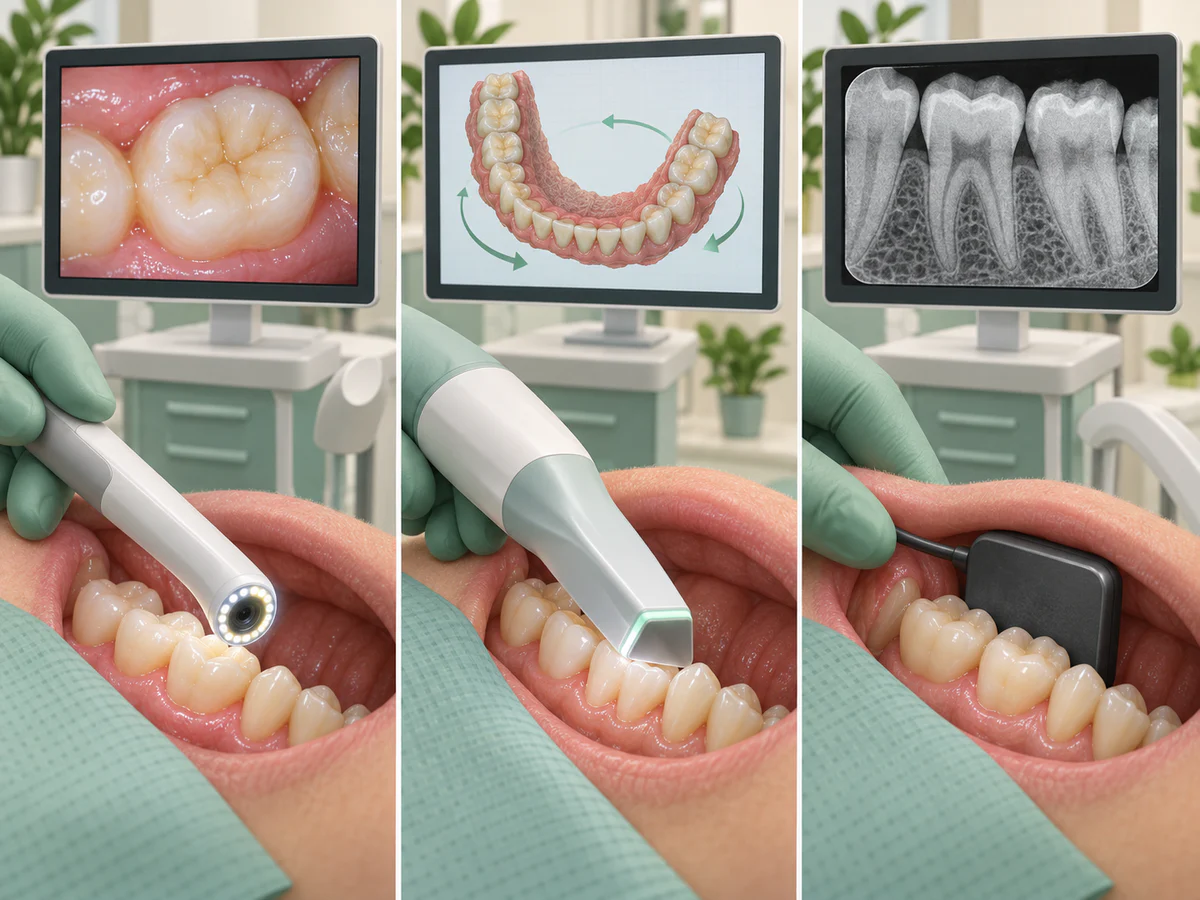
The intraoral camera should be distinguished from two related tools that are often confused with it. The intraoral scanner does more than film: it reconstructs a three-dimensional model of the dental arches, used to fabricate a prosthesis or aligners. That is a different use, described in our article on digital impressions with an intraoral scanner. X-rays, for their part, pass through the tissues to show the inside of the tooth and the bone: the camera sees only surfaces. This difference is crucial, and we return to it below.
In practice, the camera is used above all to document and show what is visible to the naked eye but hard for you to perceive, for lack of the right angle and lighting. A back tooth, the inner surface of a molar, or the edge of a gum finally become observable on a screen, right at your eye level.
Seeing to Understand: The Camera’s Real Contribution
The best-documented benefit of the intraoral camera is educational. When patients see the buildup of plaque or an inflamed gum for themselves, they better understand why a treatment or a change in habits is being proposed. The review by Pentapati and Siddiq (Clinical, Cosmetic and Investigational Dentistry, 2019) highlights precisely this use: the camera helps visualize the areas where biofilm accumulates and inflamed tissues, which can improve hygiene knowledge, attitudes, and practices and motivate patients toward appropriate methods.
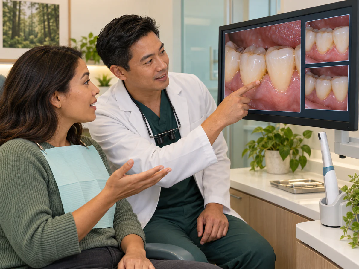
What is sometimes called “shared visual diagnosis” rests on a simple idea: we act more readily on a problem we have seen than on one that has merely been described to us. The landmark study by Willershausen and colleagues (International Dental Journal, 1999), conducted on one hundred patients split into two groups, illustrates this effect: both groups reduced their dental plaque, but the group that benefited from the camera achieved better results, and 88% of participants found the additional visual information useful and desirable.
More recent work, however, tempers the extent of this benefit. The eight-month randomized controlled trial by Araújo and colleagues (International Journal of Dental Hygiene, 2019), which compared camera use and text messaging for gingivitis control in 142 adults, shows that these aids can support hygiene behaviors, but that the long-term effect depends largely on the patient’s sustained commitment. The camera can trigger awareness; it does not, on its own, sustain motivation.
An Aid to Decision-Making and Trust
Seeing your own case changes the nature of the conversation. Rather than passively accepting a decision, the patient takes part in it: they locate the problem, gauge its extent, and understand the options. This transparency builds trust, and it is also an important point for informed consent — the agreement given after truly understanding what is being proposed.
The camera also makes follow-up easier. Comparing a photo from one visit to the next makes it possible to observe objectively an improvement in hygiene, the healing of a gum or, on the contrary, a worsening situation. For conditions in which the patient plays a central role, such as bleeding gums or periodontitis versus gingivitis, this concrete visual evidence is often worth more than a long explanation.
One pitfall should nevertheless be avoided: seeing is not understanding. A magnified image can be alarming and suggest an emergency where there is none, or on the contrary provide false reassurance. Interpretation remains the practitioner’s role — placing the image within the full examination, the patient’s history and, where relevant, the X-ray. The camera shows; it does not explain in your place.
What the Intraoral Camera Does Not Do
This is probably the most important point, and the one least emphasized in marketing materials. The camera has clear limits that you should know about, so as not to credit it with powers it does not have.
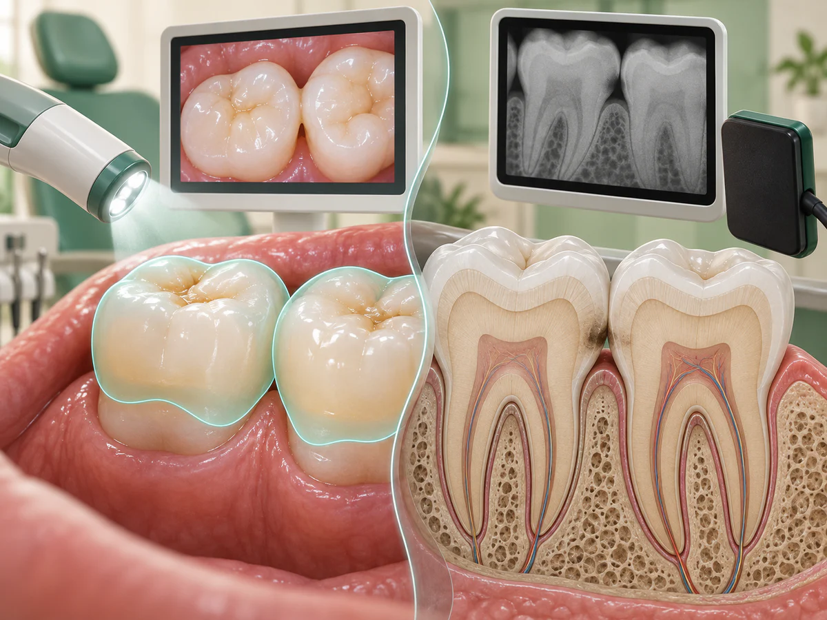
It sees only the surface. An optical camera does not pass through enamel or bone. It therefore shows neither the inside of a tooth, nor a cavity hidden between two teeth, nor an infection at the root, nor the bone that supports the teeth. Yet cavities located between the teeth frequently escape visual examination alone: this is precisely what the systematic review with meta-analysis by Schwendicke and colleagues (Journal of Dentistry, 2015) confirms, reporting low sensitivity for the detection of proximal lesions and concluding that X-rays remain the reference for identifying these interproximal cavities. The camera, which explores only visible surfaces, therefore never replaces an X-ray when one is indicated.
It does not provide a diagnosis on its own. An image, however sharp, must be interpreted. The systematic review with meta-analysis by Mohammad-Rahimi and colleagues (Journal of Dentistry, 2025), which pooled eighteen studies and around 1,300 participants, concludes that visual assessment of clinical photographs achieves clinically acceptable accuracy for detecting caries, comparable to direct visual examination, but that it complements rather than surpasses it. The camera documents what is visible well; it does not do away with the clinical examination or clinical judgment.
It does not treat, and it guarantees no outcome. Showing a problem is only a first step. The camera changes neither the indication for a treatment, nor the quality of the procedure, nor the follow-up. It can, on the other hand, give a false impression of precision: a beautiful high-definition image remains a partial, two-dimensional view of a three-dimensional reality.
| Question | What the intraoral camera provides | What it does not provide |
|---|---|---|
| Seeing the condition of tooth and gum surfaces | A magnified, live view shared with the patient | Cannot see inside the tooth or the bone |
| Detecting a cavity between two teeth | Sometimes spots a sign on the surface | Does not replace X-rays for proximal lesions |
| Understanding and deciding on a treatment | Improves understanding and motivation | Does not make the diagnosis in the practitioner’s place |
| Tracking changes over time | Allows images to be compared from one visit to the next | Guarantees neither improvement nor results |
What an Examination with a Camera Looks Like
Using the camera fits into the examination without noticeably lengthening it. The process is simple:
- the practitioner first performs the usual clinical examination;
- they introduce the camera and move it over the areas of interest, teeth and gums, while the image is displayed on the screen;
- they capture a few images of the points worth explaining or keeping in your file;
- they go over what is visible with you, distinguishing what is certain from what calls for further examination;
- if necessary, they follow up with an X-ray or another examination for what the camera cannot see.
This tool is generally painless and non-invasive: it is only a camera, with no radiation. It does not, however, replace rigorous daily hygiene, the only thing capable of preventing plaque and inflammation day after day, as detailed in our article on daily dental hygiene. The camera makes visible what we neglect; it is then up to each of us to act.
The Intraoral Camera’s Rightful Place
Used for what it is, the intraoral camera is an excellent tool for patient education and transparency. It turns a consultation into an exchange in which you see, understand, and decide, and it makes follow-up easier by objectively documenting changes. These benefits are real and documented with regard to patient understanding and motivation.
Presented poorly, it can instead suggest a complete diagnosis when it shows only surfaces, or imply a guarantee of results that no device can give. The rule is simple: the camera complements the clinical examination and, when indicated, the X-ray; it does not replace them. Its value lies in the quality of the dialogue it makes possible, not in the technology itself.
In Summary
The intraoral camera is a small camera that displays the inside of your mouth on a screen, to help you see and understand your situation. Its main contribution is educational: seeing your own problem fosters understanding, trust, and motivation, as shown by the work of Willershausen and colleagues (1999), Pentapati and Siddiq (2019), and Araújo and colleagues (2019). But it has clear limits: it sees only surfaces, does not detect cavities hidden between the teeth or internal lesions, replaces neither X-rays nor the clinical examination, and guarantees no outcome. It is a valuable communication tool, provided it is kept in its rightful place: that of an aid to decision-making, never that of a diagnosis or a treatment in itself. For any specific situation, the advice of a dental surgeon, in Kenitra or elsewhere, remains essential.
Frequently asked questions
What is an intraoral camera used for at the dentist's office?
Can the intraoral camera detect all cavities?
Does the intraoral camera replace X-rays?
Does seeing my teeth on screen really help me take better care of them?
Is an examination with an intraoral camera painful or dangerous?
Is a camera image enough to make a diagnosis?
Sources
Medical references consulted for this article.
- 1Willershausen B, Schlösser E, Ernst CP, The intra-oral camera, dental health communication and oral hygiene, International Dental Journal, 1999
- 2Pentapati KC, Siddiq H, Clinical applications of intraoral camera to increase patient compliance - current perspectives (revue), Clinical, Cosmetic and Investigational Dentistry, 2019
- 3Araújo MR, Alvarez MJ, Godinho CA, Roberto MS, An eight-month randomized controlled trial on the use of intra-oral cameras and text messages for gingivitis control among adults, International Journal of Dental Hygiene, 2019
- 4Mohammad-Rahimi H et al., Accuracy of clinical photography for the detection of dental caries (revue systématique et méta-analyse), Journal of Dentistry, 2025
- 5Schwendicke F, Tzschoppe M, Paris S, Radiographic caries detection: a systematic review and meta-analysis, Journal of Dentistry, 2015
- 6Fernández CE, Maturana CA, Coloma SI, Carrasco-Labra A, Giacaman RA, Teledentistry and mHealth for Promotion and Prevention of Oral Health (revue systématique et méta-analyse), Journal of Dental Research, 2021
Contents
Share