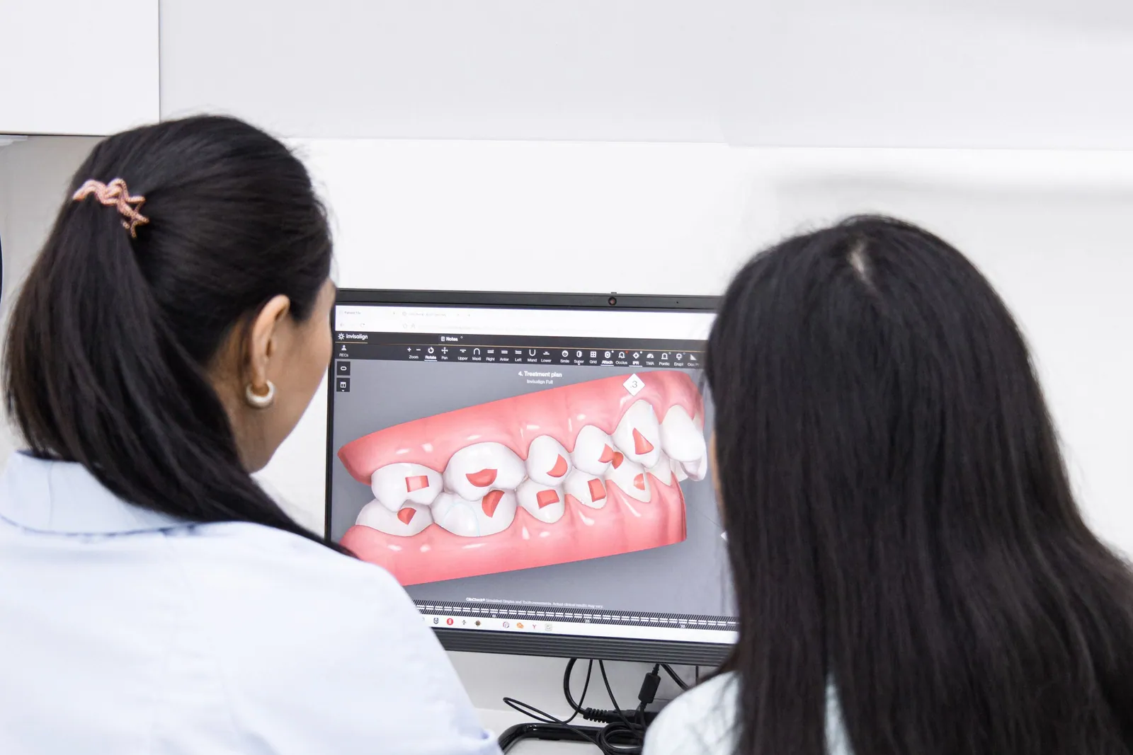
The “fully digital workflow” describes a way of designing a dental treatment plan in which the patient’s data — the shape of the teeth, the bone, and sometimes the face — is captured, assembled and planned on a computer, from the very first examination through to the treatment proposal.
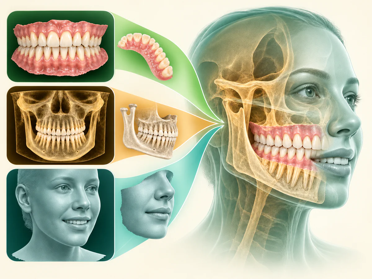
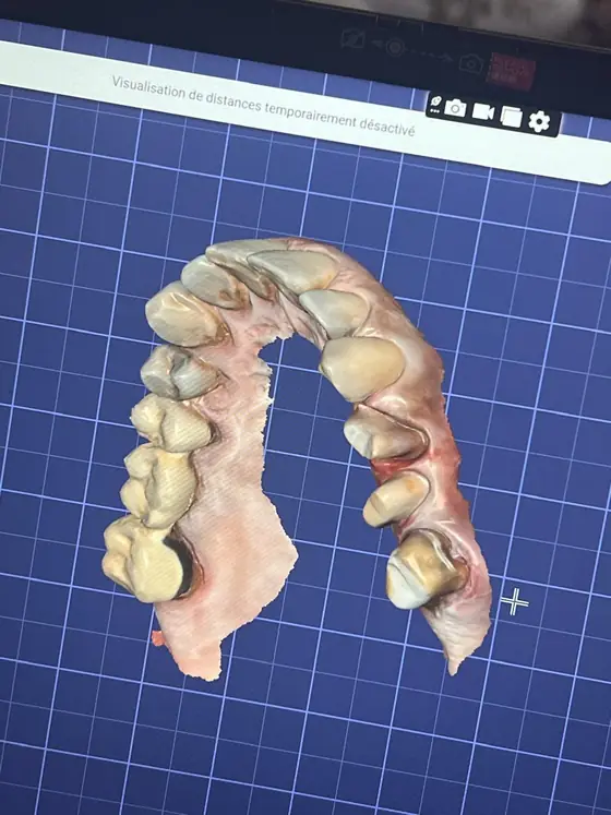
At the heart of this approach lies an appealing idea: merging three very different types of images — the optical impression of the teeth (intraoral scan), 3D imaging of the bone (CBCT) and three-dimensional photography of the face (facial scan) — into a single, coherent 3D model.
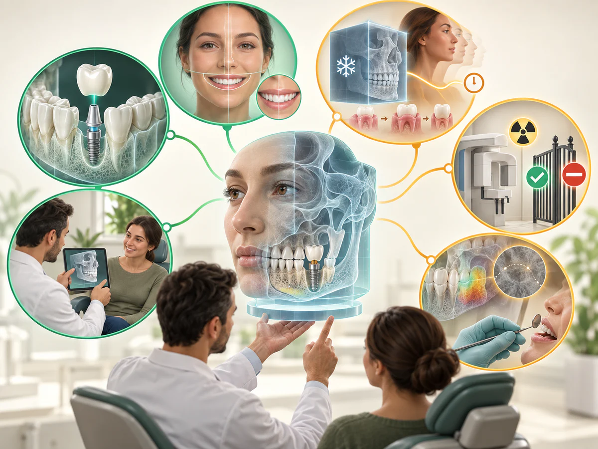
This model, sometimes called the “virtual patient” or the patient’s “digital twin”, aims to represent both function (the occlusion, the available bone) and esthetics (the smile within the face), so that treatment can be planned in a more global and more personalized way.
This article is an educational explainer about the technology in general, and not a description of the equipment found in any particular practice.
As a dental surgeon in Kénitra, I offer a measured reading of it here: what a digital workflow and a digital twin really are, how the images are merged, what research says about their accuracy and their usefulness, and — this last point is essential — what this technology does not provide.
Every statement points to a verifiable source, and the figures quoted are study data, to be read as orders of magnitude, never as a guarantee for any individual case.
What is a digital workflow and a “digital twin”?
A workflow is described as digital when the steps that are usually manual — the paste impression, the plaster model, planning on a 2D radiograph — are replaced by digital data processed in software. The workflow may be partial (only the optical impression, for example) or move towards being “fully digital”, where capture, planning and sometimes fabrication follow on from one another with no analog step in between.
The “digital twin” (or virtual patient) is the conceptual culmination of this: a single 3D model bringing together several sources of data. As Park and colleagues describe it (Journal of Craniofacial Surgery, 2022), the idea is to “integrate CBCT, the intraoral scan and the 3D facial scan” in order to obtain a model that can serve for the initial consultation, for diagnosis, for planning, for treatment simulation and for evaluating results.
It is therefore not one more examination, but a way of assembling examinations that already exist so they can be looked at together.
One point of vocabulary needs to be underlined straight away. The term “digital twin” is borrowed from industry, where it refers to a virtual replica updated in real time. In dentistry we are still a long way from that: as things stand, the model is a frozen 3D photograph of the patient at one given moment, not a living, dynamic copy. It is a tool for representation and planning, not an autonomous double.
The three data sources: what each one contributes
The strength of the model comes from the way three separate acquisitions complement one another, because none of them captures everything on its own.
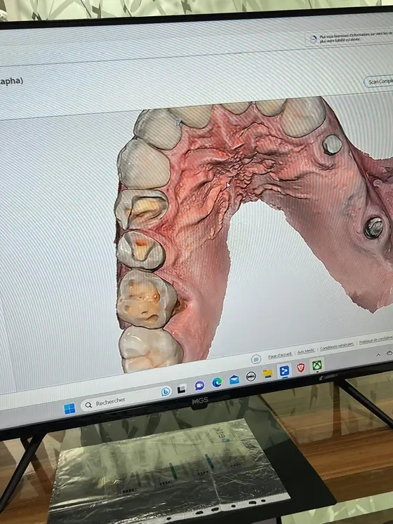
- The intraoral scan records the surface of the teeth and gums in fine detail, along with the occlusion, and produces a digital impression. Its indications and its limits are set out in our article on the digital impression with an intraoral scanner. On the other hand, it sees neither the bone, nor the roots, nor the face.
- The CBCT (cone beam) provides 3D imaging of the bone, the roots and the deeper structures, which is essential in implantology in particular. Its principle, its indications and its benefit–risk balance are explained in our article on CBCT and 3D dental imaging. But the CBCT renders the fine surface of the teeth and the texture of the facial soft tissues poorly.
- The facial scan captures the shape and surface of the face in three dimensions, which makes it possible to place the future smile back into its esthetic context. It is the most recent addition, and the most technically demanding.
It is precisely because these three sources are individually incomplete that the idea of superimposing them is interesting: the intraoral scan for dental detail, the CBCT for the bone, the facial scan for the face. Brought together, they sketch out a complete patient, from the bone to the smile.
How are the images merged?
The fusion relies on an operation known as superimposition (or registration): the software seeks to align common points or surfaces across the different files so as to position them correctly in relation to one another. In practice, the imaging files (DICOM, from the CBCT) and the surface files (STL, from the scanners) are imported into planning software, which matches them up.
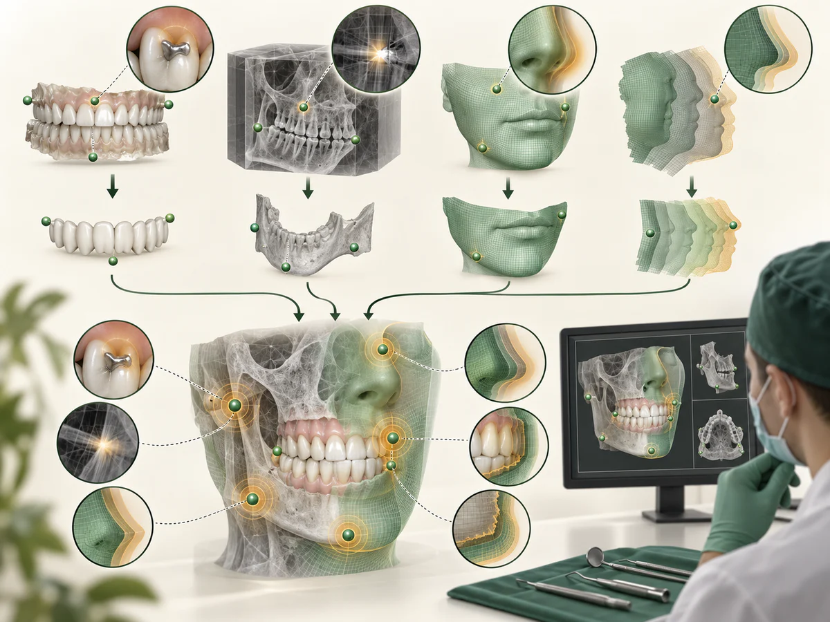
The difficulty is real. Shujaat and colleagues (Dentomaxillofacial Radiology, 2021), in a review of the integration of imaging modalities, underline a central principle: “the accumulation of errors at each step of the chain can negatively influence the final outcome”. In other words, every acquisition and every alignment introduces a small imprecision, and these imprecisions add up.
The authors detail several sources of error: metal artifacts (crowns, appliances) that disturb how the CBCT is read, the difficulty the CBCT has in faithfully representing the soft tissues of the face, and the time gap between the CBCT acquisition and that of the face. Perfect registration, they conclude, remains difficult to achieve.
To limit the error, some protocols use common landmarks: extraoral and intraoral “scan bodies” acting as anchor points. Schobben and colleagues (Clinical Oral Investigations, 2025) compared two non-irradiating methods for integrating an intraoral scan into a 3D facial photograph: depending on the method, the deviation from the reference varied, and not all measurements yet reached the clinically acceptable threshold.
This result illustrates the state of the art well: the fusion works, but its accuracy depends on the method and remains open to improvement.
What is a digital twin used for in practice?
The value of a unified model can be found at several levels, provided one stays realistic about what it makes possible.
First, implant planning. Having the bone (CBCT) and the projected restoration (intraoral scan) within the same model makes it possible to plan the implant position according to the future crown, rather than the other way round.
A systematic review by Saini and colleagues (Digital Health, 2024), covering 21 studies, concludes that virtual planning on CBCT data and the surgical guides derived from it show greater accuracy than traditional methods, with mean deviations at the entry point of around 1 mm and angular deviations of approximately 3.8° on average. This subject is developed in our article on guided implant surgery.
Next, esthetic planning. By placing the smile project back into the face, the facial scan feeds into an esthetic design approach. A systematic review and meta-analysis by Saini and colleagues (Digital Health, 2025) on artificial-intelligence-assisted digital smile design observed an improvement in measurable parameters — smile symmetry, position of the lip arch, visibility of the incisal edges — while also noting strong heterogeneity between studies and a lack of data on long-term durability.
Finally, communication. The literature converges on one point: the 3D model improves the dialogue with the patient and the acceptance of the treatment plan, by making the project visible before it is carried out. That is a real benefit, but one that belongs to patient education and to the predictability of the project, not to any guarantee of a clinical result.
| Data source | What it captures | Its main limitation |
|---|---|---|
| Intraoral scan | Surface of the teeth, gums, occlusion | Sees neither the bone, nor the roots, nor the face |
| CBCT (cone beam) | Bone, roots, deep structures (3D) | Soft tissues and fine tooth surface poorly rendered; X-rays |
| 3D facial scan | Shape and surface of the face | Demanding acquisition; delicate alignment |
| Digital twin (fusion) | Overall view of function + esthetics | Errors from each step accumulate |
What the digital workflow does not provide
Honest information means clearly naming the limits of this approach, because the term “digital twin” can give the impression of absolute accuracy.
First, a model is neither a diagnosis nor a guarantee. Superimposing images is a decision-support tool: it replaces neither the clinical examination, nor experience, nor the practitioner’s judgement. A fine-looking 3D model may rest on an imperfect alignment and prove misleading if it is taken at face value.
Second, accuracy has a ceiling, and errors add up. As Shujaat and colleagues (2021) point out, every link in the chain — acquisition, segmentation, registration — introduces a margin of error, and the final result accumulates those margins. Metal artifacts, patient movement or a time gap between examinations all degrade the fusion. The work of Schobben and colleagues (2025) shows that, depending on the technique, not all measurements yet reach the clinically acceptable threshold.
Third, the model is frozen, not living. Unlike an industrial digital twin, the dental model remains an image at one given moment; it does not update itself with healing, ageing or real functional movement.
Fourth, the CBCT remains an irradiating examination, to be used only where the indication is justified, as the logic of the ALARA principle reminds us. Multiplying acquisitions in the name of “going fully digital” is not justified: the decision to image must rest on an expected benefit, not on the appeal of the technology.
Finally, the evidence remains young and heterogeneous. The reviews cited almost all underline the same reservations: small numbers, variable methodologies, limited follow-up, a lack of data on durability and on cost-effectiveness. The digital workflow is a promising field, advancing quickly, not a mature technology whose benefits have all been demonstrated over the long term.
In summary
The fully digital workflow consists of capturing, merging and planning the patient’s data on a computer, to the point of building a “digital twin” that brings together the intraoral scan (the teeth), the CBCT (the bone) and the facial scan (the face) in a single 3D model (Park et al., 2022).
This integration has real benefits: more accurate implant planning through surgical guides (Saini et al., 2024), esthetic design better placed within the face (Saini et al., 2025), and better communication with the patient.
But it also has clear limits: the accumulation of errors at each step of the chain (Shujaat et al., 2021), a fusion whose accuracy depends on the method and remains open to improvement (Schobben et al., 2025), a model that is frozen rather than living, the irradiating nature of the CBCT, and a level of evidence that is still young.
The digital twin is a tool serving diagnosis, planning and dialogue — not a substitute for clinical judgement, nor a promise of results.
Frequently asked questions
What is a patient’s “digital twin” in dentistry?
How are the intraoral scan, the CBCT and the facial scan merged?
Does the digital workflow make planning more accurate?
Does the digital twin guarantee the esthetic result?
Does going fully digital mean more radiographs?
What are the main limits of this technology?
Sources
Medical references consulted for this article.
- 1Park JH, Lee GH, Moon DN, Yun KD, Kim JC, Lee KC, Creation of Digital Virtual Patient by Integrating CBCT, Intraoral Scan, 3D Facial Scan: An Approach to Methodology for Integration Accuracy, Journal of Craniofacial Surgery, 2022
- 2Shujaat S, Bornstein MM, Price JB, Jacobs R, Integration of imaging modalities in digital dental workflows — possibilities, limitations, and potential future developments, Dentomaxillofacial Radiology, 2021
- 3Schobben RRP et al., Two experimental methods to integrate intra-oral scans into 3D stereophotogrammetric facial images, Clinical Oral Investigations, 2025
- 4Saini RS et al., Impact of 3D imaging techniques and virtual patients on the accuracy of planning and surgical placement of dental implants (revue systématique), Digital Health, 2024
- 5Saini RS et al., Impact of artificial intelligence-based digital smile design on patient and clinician satisfaction and facial esthetic outcomes (revue systématique et méta-analyse), Digital Health, 2025
- 6Burlacu Vatamanu OE, Cristache CM, Drafta S, Nimigean VR, Evaluation of Four 3D Facial Scanning Technologies: From Photogrammetry to Structured-Light Systems in Clinical Dentistry, Dentistry Journal, 2026
Contents
Share